Stem Cell Blog
Употребата на матичните клетки од папочна врвца рапидно се зголемува. Пред 10 години крвта од папочна врвца можеше да лекува околу 40 состојби, но денес таа бројка е над 80. Со нетрпение очекуваме нови терапии за болести и нарушувања како што се дијабет, аутизам и мозочен удар, можете да бидете во тек со најновите случувања во регенеративната медицина на нашиот блог за матични клетки.

A new study published in the journal Burns & Trauma has found that the application of stem cells promotes the repair of diabetic wounds. [1]
Affecting nearly 1 in 10 adults worldwide, diabetes is quickly becoming one of the most prevalent and widespread causes of global public health concern. [2]
Along with causing high blood sugar levels, diabetes can lead to comorbidities that can drastically affect and complicate diabetes sufferers’ quality of life and health.
Chronic wounds, such as diabetic foot ulcers, are the leading cause of such complications, which can result in disability and even, in some cases, mortality.
Diabetic wounds heal more slowly because normal cellular processes are interrupted by high glucose levels.
One important cellular process that high glucose particularly affects is autophagy, the process by which damaged cells are broken down by the body and recycled towards other areas of cellular repair. Autophagy plays a pivotal role in the healing process. [3]
What the team at Shengjing Hospital of China Medical University found was that the application of exosomes from adipose-derived mesenchymal stem cells significantly improved the healing process of wounds in diabetic mice.
They observed the exosomes upregulating the autophagy flux, meaning increased epidermal cell proliferation and migration, promoting healing.
These findings are highly promising, suggesting that mesenchymal stem cell derived exosomes have the potential to reduce the risks posed by diabetic wounds and speed up the recovery process for those who suffer from them.
Currently 50%-70% of all amputations are for diabetic foot ulcers. Depending on further findings, the use of stem cells in the treatment of diabetic wounds like foot ulcers may even mean an end for drastic last resort measures, such as amputation. [4]
If you want to know more about the potential of stem cells and about how to privately store the mesenchymal stem cells from your baby’s umbilical cord and placenta, download our FREE Welcome Pack below.
References

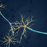
There’s exciting news from the University of Galway, where researchers are developing a new technique that they hope will improve the viability of stem cell treatments for Parkinson’s disease.
The pioneering method, which involves the use of a substance called hydrogel, could revolutionise the way stem cell treatments for Parkinson’s disease are carried out, making them more effective and less prone to failure.
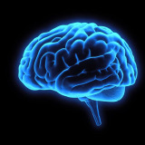
Parkinson’s disease is a neurological condition affecting around 150,000 people in the UK. [1]
The condition, which is caused by a lack of dopamine in the brain, manifests in severe ways with symptoms including tremors, muscle rigidity and slowness of movement. [2]
Unfortunately, Parkinson’s is degenerative, meaning that it gets worse over time. It can make people who live with the condition more vulnerable to poor health and disability, which can end up having fatal repercussions in some cases.
The degeneration of nerve cells in the brain is what results in the underproduction of dopamine. The aim of recent stem cell research has been to find a way of repairing and replacing these cells through the use of induced stem cells.
These induced stem cells are harvested from different areas of the body, such as skin, and then reprogrammed to become the type of cells necessary for brain repair.
However, these cells require transplantation at a very early stage of their development into brain cells and once transplanted, many of them do not end up converting.
What researchers at the University of Galway have discovered is that by transplanting these induced stem cells in a collagen hydrogel, effectively a water-based scaffold, significantly improves the chances of the stem cells both surviving and then differentiating into the cells necessary for therapy. [3]
With funding from the Michael J. Fox Foundation for Parkinson’s Research (MJFF), the study’s findings were published in the Journal of Neural Engineering and have been met with widespread acclaim.
The research is ongoing, but the team at the University of Galway and MJFF hope that this new transplantation technique will significantly improve outcomes for sufferers of Parkinson’s disease.
If you want to learn more about how you could give your baby access to future stem cell therapies, download our FREE Welcome Pack below.
References

02/04/2024 BlogStem Cell NewsStem Cell Therapies
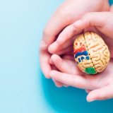
This week it was announced that researchers have grown functioning ‘mini-organs’ using the stem cells in amniotic fluid in what’s being hailed as a huge breakthrough in the world of prenatal medicine.
Scientists at University College London and Great Ormond Street harvested the discarded stem cells from the amniotic fluid of 12 foetuses. [1]
The team were then able to grow what’s known as ‘organoids’ from these stem cells, which are effectively small replicas of real human organs. [2]
These ‘organoids’ have huge potential in clinical trials for new and experimental medicines, providing researchers with a window into understanding how healthy organs function compared to diseased ones.
The stem cells the UCL and GOSH team harvested from the amniotic fluid were the progenitor cells for lungs, kidneys and intestines. Because these stem cells, discarded during pregnancy by the foetus as is normal during pregnancy, are tissue specific they contain information necessary for the development and functionality of particular organs.
How can these organoids be used?
Collaborating with a team from KU Leuven in Belgium, the researchers studied the development of babies with CDH, otherwise known as congenital diaphragmatic hernia, a condition that affects around 1 in 5,000 babies every year. [3]
CDH is a disease that causes a hole in the diaphragm of the developing foetus, causing organs like their liver and intestines to rise and interfere with the capacity of the lungs.
Around 7 in 10 of babies born with CDH survive, and once the hernia is identified doctors can even operate on foetuses whilst they’re still in the womb. [4]
The team used organoids grown from the stem cells of healthy babies and babies affected by CDH both before and after treatment in order to determine whether there were any important differences between the biological characteristics of their organs.
What the team found was that there were significant developmental differences between the organoids that were healthy and the organoids that were affected by CDH.
By using these stem cell grown organoids researchers were able to determine that the organ progenitor cells taken from the amniotic fluid of babies affected by CDH themselves exhibit features of the disease.
Despite this stem cell-based research being in the very early stages of development, studies like this one show just how powerful stem cells are and just how important they’ll be for the future of medicine.
If you’d like to learn about banking your baby’s stem cells so that they have access to the future of medicine, sign up to download your free welcome pack below.
References

Therapies involving stem cells hold immense promise for treating a variety of medical conditions, particularly spinal cord injuries.
A future where damaged spinal cords can be regenerated through the medical application of stem cells, restoring mobility and function to those affected, isn’t as far away as it once seemed.
Especially since scientists at MIT and the Singapore-MIT Alliance for Research and Technology have taken a significant step towards this future by developing a tiny device that could enhance the safety and effectiveness of stem cell treatments.
Understanding Cell Therapy
Stem cell therapy involves reprogramming the stem cells taken from a patient’s skin or blood cells to create induced pluripotent stem cells (iPSCs).
These iPSCs are then coaxed into becoming progenitor cells, specialised to differentiate into spinal cord cells.
Once these progenitor cells are transplanted back into the patient, they can regenerate part of the injured spinal cord, offering hope for recovery.
However, undifferentiated iPSCs pose a risk of forming tumours, limiting the therapy’s safety and efficacy.
Introducing the Microfluidic Cell Sorter
To address this challenge, researchers have developed what’s known as a microfluidic cell sorter.
Effectively a kind of sieve, this device is capable of removing undifferentiated cells from a batch without harming fully-formed progenitor cells.
It can sort over 3 million cells per minute and can be scaled up by chaining multiple devices together, potentially sorting more than 500 million cells per minute.
Moreover, the plastic chip housing the sorter can be mass-produced at low cost, making widespread implementation feasible.
How It Works
The sorter operates based on the size difference between residual, undifferentiated pluripotent stem cells and progenitor cells.
Pluripotent stem cells tend to be larger due to the presence of numerous active genes in their nuclei.
As cells pass through microfluidic channels at high speeds, centrifugal forces focus them at specific points, enabling their separation based on size.
By running the sorter twice at different speeds, researchers effectively remove larger cells that are associated with a higher tumour risk.
Promising Results and Future Directions
While the sorter doesn’t eliminate 100% of undifferentiated cells, it significantly reduces the risk, enhancing the safety of cell therapy treatments.
Further studies are underway to validate these findings in larger-scale experiments and animal models. If successful, purified cells could offer improved efficacy and safety in vivo, paving the way for broader applications of this technique.
The development of this microfluidic cell sorter represents a significant advancement in the field of stem cell therapy.
By enhancing safety and effectiveness, it brings us closer to realising the full potential of regenerative medicine for conditions like spinal cord injuries.
With ongoing research and technological innovations, the future holds promising possibilities for improving healthcare outcomes through cell-based therapies.
To find out more about how you could give your baby access to the future of medicine by banking their stem cell rich umbilical cord and placenta, download your FREE Parents Guide to Cord Blood Banking below.
Sources

It was announced this week that researchers at the University of East London had managed to grow a “heart” using stem cells.
This breakthrough could be instrumental in paving the way for life-saving research into heart disease treatments, with stem cells playing a pivotal role in the future of cardiovascular research.
According to the British Heart Foundation, there are over 170,000 deaths linked to heart and circulatory diseases each year. That’s 480 each day, or 1 every 3 minutes.
With 7.6 million people in the UK living with heart and circulatory diseases, progress in the field of cardiovascular medicine can’t come soon enough. [1]
Because the grown stem cell “heart” has the same characteristics as a normal human heart, scientists are hoping that it can be a more ethical, accurate alternative to the use of animal specimens in research.
This is the latest development in a long line of investigations into the application of stem cells to grow heart tissue.
Last April, researchers at the Francis Crick Institute and Imperial College London set about evaluating how human pluripotent stem cells (hPSCs) could be used to grow left ventricular heart muscle cells. [2]
Their findings suggested that, with the right environment, stem cells are able to differentiate successfully into the cells that make up the left ventricle of the heart, the area most commonly affected by heart disease [3]
In August of last year, a team of researchers from various Israeli institutions had grown a small, yet complete and beating, model of a heart using stem cells. They were even able to fit sensors to the model to monitor its behaviour. [4]
These so-called organoid hearts, grown from stem cells, are incredibly useful in finding treatments and therapies for cardiovascular diseases because of their likeness to human hearts.
Although these “grown” hearts are a lot smaller than human hearts, their construction and makeup reflects the different chambers, tissues and cells that make up a human heart far more accurately. This makes them better in experiments for developing medicine for heart disease than animal samples.
The team at The University of East London are also looking to develop an AI in conjunction with the stem cell heart to monitor intricate changes to cells that could indicate the onset of heart disease. [5]
With thousands of clinical trials currently underway for the application of cord blood and perinatal stem cells in regenerative medicine, it’s possible that your baby’s umbilical cord and placenta hold the key to unlocking their access to the treatments of the future.
Find out more about how storing your baby’s stem cells could safeguard their health for life by downloading our FREE Welcome Pack below.
Sources

World Cancer Day takes place this year on 4th February. A day of global unity, dedicated to raising awareness about cancer, World Cancer Day also plays an important part in dispelling myths about cancer, in addition to promoting early detection and prevention.
This year’s World Cancer Day theme is ‘Close the Care Gap’, referring to the gap between the level of care received by privileged vs underprivileged cancer patients.
On this significant day, we’re invited not only to reflect on the impact of cancer but also asked to consider how care outcomes for a cancer diagnosis could be improved.
One such way is through cord blood banking, which is already being used in over 80 treatments, including for leukaemia.
In this blog, we will delve into the importance of cord blood banking and how it is offering hope in the fight against cancer.
Understanding Cord Blood Banking
Cord blood banking involves the collection and preservation of the residual blood from the newborn umbilical cord following birth.
This precious resource contains powerful stem cells that can develop into various specialised cells, such as the ones in hair, skin, organs, blood and the nervous system.
Their unique abilities to self-replicate and differentiate positions them at the forefront of regenerative medicine, a branch of medicine that makes use of stem cells’ potential capacity to repair, renew and regrow cells and tissues to treat a range of diseases. [1]
The Benefits of Cord Blood Banking
Cord blood stem cells are the approved therapy for over 80 diseases, including leukaemia, neuroblasts, and certain genetic disorders.
Their relative naivety and plasticity when compared to stem cells derived from other sources makes them some of the purest and most powerful forms of stem cell available.
Cord blood stem cells are a 100% match for your baby, meaning that they can be used in therapies without risk of rejection. They also have a good chance of being a perfect match for siblings and a partial match for family members, offering a safer and more accessible option for transplantation.
A painless and non-invasive procedure, cord blood collection is safe, non-invasive and poses little to no risk to the mother or baby.
As medical research advances, the potential uses of cord blood stem cells continue to expand.
Researchers are exploring their use in regenerative therapies that seek to harness the power of these stem cells in order to combat diseases that are currently incurable, including some forms of cancer. [2]
Promoting Cord Blood Banking on World Cancer Day
World Cancer Day provides an ideal platform to educate expectant parents and the general public about some of the most pioneering research happening to combat cancer, amongst them: cord blood banking.
Understanding the potential lifesaving impact of this resource is the first step in motivating more families to consider this option.
World Cancer Day reminds us of the global challenge posed by this disease, but it also presents an opportunity to promote hope and innovative solutions.
Cord blood banking is one such solution that has the potential to save lives and help in the battle against cancer.
Sarah’s Story
For eight year old Sarah, for instance, cord blood banking was the last hope she had after both chemotherapy and a bone marrow transplant proved ineffective in the treatment of her acute myeloid leukaemia. [3]
A form of cancer that attacks the monocyte or granulocyte cells, naive progenitor white blood cells from bone marrow, acute myeloid leukaemia predominantly affects children and young people.
Chances of a full recovery are rarely good.
Having undergone a bone marrow transplant from her brother, Sarah initially showed promising signs of recovery, until the cancer returned.
Rounds of emergency chemotherapy were required to try to keep the cancer at bay, but it continued to return.
Seeing no other option for Sarah, doctors at the Royal Manchester Children’s Hospital offered her a pioneering stem cell transplant using donated cord blood.
Incredibly, thanks to this treatment Sarah and five other children who also participated in the trial, are now in remission; their access to a healthy, happy life restored to them.
Although this transplant was the result of donation, privately banking cord blood stem cells means that your baby always has access to their own perfect donor match: themselves.
This drastically reduces the risk of rejection should they ever need to access a therapy in future like Sarah’s.
With thousands of clinical trials currently underway to explore the potential uses for umbilical cord blood stem cells in a range of regenerative treatments, storing these precious cells the day baby is born could safeguard their future for years to come.
For more information about the power of cord blood banking, download your FREE Welcome Pack below.
Sources

02/02/2024 BlogStem Cell NewsStem Cell Therapies
This month sees two important missions lifting-off into space, both of which will carry out pioneering stem cell research.
The first of these has already launched. On 18 January, Ax-3, the third mission by private company Axiom to be launched using Space X rockets, departed for the International Space Station. [1]
Aboard were four astronauts who over the course of two weeks will carry out a variety of experiments in microgravity.
Amongst them will be several significant ones relating to stem cells.
The Sanford Stem Cell Institute (SSCI) operating out of the University of California, San Diego will be paying close attention to the astronauts’ findings in relation to their investigation into tumour organoids. [2] [3]
By analysing the growth rates of cancer stem cells, SSCI hope to build on previous Axiom missions and shed light on how cancer develops in order to identify early warning signs.
Aboard the same mission, the National Stem Cell Foundation (NSCF) are seeking to utilise 3D brain models to help ascertain key onset markers of neurodegenerative diseases. [4]
NSCF Researchers hope that by analysing the effects of microgravity on the 3D models, which are derived from the induced pluripotent stem cells of patients with either Parkinson’s or primary progressive multiple sclerosis, they’ll be able to better understand how these diseases develop.
In this, the third Axiom mission of its kind, researchers hope to take advantage of the different rates at which stem cells develop in space to point out what the future of treatment looks like for some of the most harmful diseases on Earth.
The second significant space mission to launch this month will be on the 29 January.
This mission, which has as its main payload materials for the resupply of the International Space Station, will investigate how the absence of gravity plays a role in bone loss. [5]
A team from the Mayo Clinic in Florida will analyse the effect of gravity, or lack thereof, on mesenchymal stem cells derived from bone marrow.
Their findings could end up having an impact on the course of clinical trials for a variety of conditions, such as osteoporosis, that take advantage of the powerful regenerative potential of these type of stem cells to regrow bone tissue.
This mission will be the first of two, with the next scheduled tentatively for the end of the year. This second flight should seek to investigate the effect of microgravity on different cell types that similarly occasion either bone formation or loss.
These experiments in space show the truly pioneering research taking place in the field and illustrate the huge potential these incredible cells have for humans on Earth now, and possibly in space in the near future.
Find out more about what stem cells can do by requesting your FREE Welcome Pack below.
Sources
[1] Ax-3 Mission Research. Axiom Space. https://www.axiomspace.com/missions/ax3/research

22/01/2024 BlogStem Cell NewsStem Cell Therapies
Although hair loss is common, it can have a huge impact on a person’s self-esteem and, subsequently, their physical and mental wellbeing.
New trials exploring the potential applications of stem cells in treating hair loss offer hope for those who are suffering both now and in the future.

The first phase of a clinical trial hoping to treat Multiple sclerosis (MS) with stem cells has yielded promising results.
The early-stage trial was conducted by researchers at the University of Cambridge, along with scientists from the University of Milan Bicocca and La Casa Sollievo della Sofferenza hospital in Italy. [1]
Cells4Life Group LLP,
Units 2-3 Oak House, Woodlands Office Park,
Albert Drive, Burgess Hill, RH15 9TN, UK.
Med Bio Genetics
Pero Nakov 110, Skopje, Republic Macedonia
+389 70 221 602
info@cells4life.mk
Cells4Life Blog Posts

12/02/2026