Stem Cell Blog

Употребата на матичните клетки од папочна врвца рапидно се зголемува. Пред 10 години крвта од папочна врвца можеше да лекува околу 40 состојби, но денес таа бројка е над 80. Со нетрпение очекуваме нови терапии за болести и нарушувања како што се дијабет, аутизам и мозочен удар, можете да бидете во тек со најновите случувања во регенеративната медицина на нашиот блог за матични клетки.


sun_damage-980x842-1.jpg

A recent study, carried out both in vitro and in vivo on a mouse model, has found that extracellular vesicles derived from adipose (fat) and umbilical cord mesenchymal stem cells could have a regenerative effect on sun-damaged skin.[1]

Sun damage causes and consequences

Unprotected exposure to harmful ultraviolet (UV) rays from the sun damages the DNA in your skin, ageing it prematurely. This is called photoaging, or sun damage, and can take several forms:[2] [3] [4]

  • Wrinkles and skin thickening: UV rays break down collagen and elastin, proteins which give your skin its structure and elasticity. As a result, skin becomes thicker and wrinkled over time, beyond what would normally be caused by ageing.

  • Sun spots, redness or blotchiness: skin cells produce melanin as a reaction to UV rays, in an attempt to protect against further sun damage. This is the same process that gives you a tan. However, over time, the damaged skin becomes permanently, unevenly pigmented, causing darker patches often called sun spots. This can also take the form of broken blood vessels, causing permanent redness or blotchiness. This happens particularly in areas frequently exposed to the sun, such as the face and hands.

  • Skin cancer: too much UV exposure can cause skin cancer. In the UK, overexposure to UV radiation is the cause of 85% of cases of melanoma skin cancers.[5]

What are extracellular vesicles? 

Extracellular vesicles are tiny particles which are generated from cells. They can carry important biomolecules, like fats and proteins, to other cells, working as a messenger of sorts.

Because the contents of extracellular vesicles can vary depending on the originating cells, scientists believe they could be of great importance in medicine. They could serve as a prognostic tool to predict the likely course of diseases, as well as help to cure them.[6]

In particular, extracellular vesicles derived from mesenchymal stem cells could inherit their regenerative properties, and have been a recent focus of research for their therapeutic potential.

What did the study find?

In the study, scientists from Peking Union Medical College, Beijing, China, aimed to investigate the effects of extracellular vesicles derived from adipose mesenchymal stem cells (AMSC-EVs) and umbilical cord mesenchymal stem cells (HUMSC-EVs) on photoaging.

In vitro, AMSC-EVs and HUMSC-EVs had positive effects on keratinocytes (cells which make up the outermost layer of the skin) and fibroblasts (cells which make up the connective tissue in the middle layer of the skin) that had been exposed to UV radiation. Treatment with EVs lowered inflammation and reduced the levels at which various biomarkers of senescence (ageing) were present in cells. Moreover, the treatment boosted cell proliferation and migration, properties which make skin cells better able to heal damage. Similar protective and regenerative effects were also observed using an in-vitro, full-thickness model of human skin.

These positive results prompted further analysis in vivo, using nude mice. The mice were randomly split into four groups, keeping one as the control. The other three groups were first exposed to high doses of UV radiation, then treated, respectively, with phosphate-buffered saline (PBS) as a placebo, with AMSC-EVs and with HUMSC-EVs.

All three groups exposed to UV initially developed deep, wide wrinkles. By the end of the observation period, however, the skin of mice treated with EV showed significantly fewer and thinner wrinkles. Skin analysis showed that EV treatment helped the skin recover water content, and reversed the epidermal thickening caused by UV radiation. The treatment also improved collagen and elastin levels and reduced inflammation. Additionally, there were fewer biomarkers of ageing in the skin cells of treated mice.

The benefits of stem cell banking

Both types of EVs studied proved effective in mitigating photoaging. However, the researchers noted that, in the mouse model, the effects of EVs derived from umbilical cord MSCs seemed better, from a therapeutic point of view, than those of EVs derived from fat tissue MSCs. The skin of the mice in the HUMSC-EV treatment group was noticeably less wrinkled. Additionally, the skin’s water content was much closer to that of the mice who had not been exposed to UV radiation at all, as was the epidermal thickness.

Furthermore, the process of collecting fat tissue for therapies is inherently invasive, but must be undergone if patients wish to use autologous (their own) stem cells as a therapeutic source. Conversely, the collection of stem cells from the umbilical cord is a painless, entirely non-invasive process; however, the cord must have been collected immediately after birth, and the cells and tissues cryogenically stored for future use. If this was not done, the only way to access therapies based on umbilical cord stem cells is the use of allogeneic (donor) cords – something which can encounter any number of issues, from lack of availability to incompatibility or rejection.

To find out more about storing your baby’s umbilical cord stem cells, so they will have them ready and waiting rather than needing to seek out alternative stem cell sources should they ever need regenerative therapies, fill in the form below to request your welcome pack.

References

[1] Zhang, H., et al. (2024). Human adipose and umbilical cord mesenchymal stem cell-derived extracellular vesicles mitigate photoaging via TIMP1/Notch1. Signal Transduction and Targeted Therapy, 9(1). doi:https://doi.org/10.1038/s41392-024-01993-z

[2] Grabel, A. (2019). Photoaging: What You Need to Know About the Other Kind of Aging. The Skin Cancer Foundation. https://www.skincancer.org/blog/photoaging-what-you-need-to-know/

[3] Cleveland Clinic (2022). Sun-damaged Skin: Photoaging, Signs, Causes & Treatment.  https://my.clevelandclinic.org/health/diseases/5240-sun-damage-protecting-yourself

[4] Yale Medicine (2023). Photoaging (Sun Damage). https://www.yalemedicine.org/conditions/sun-damage

[5] Cancer Research UK (2020). Risks and causes of melanoma skin cancer. https://www.cancerresearchuk.org/about-cancer/melanoma/risks-causes

[6] Zhang, Y., Liu, Y., Liu, H. and Tang, W.H. (2019). Exosomes: biogenesis, biologic function and clinical potential. Cell & Bioscience, 9(1). doi:https://doi.org/10.1186/s13578-019-0282-2


cord_blood_leukaemia_treatment-980x980-1.jpg

After two planned transplants from unrelated donors fell through at the very last minute, Australian three-year-old Tommy Bacon is now in remission from a rare, dangerous form of leukaemia following a transplant of the stem cells from his baby sister’s cord blood.[1]

Tommy’s story 

Tommy fell ill not long after his parents discovered they were expecting a second child – a baby girl. When he first started showing signs of illness, his parents and their doctor were not immediately alarmed. They assumed it was just a case of the usual germs picked up at daycare, which he had recently begun attending.

After he developed tonsillitis during a family trip to the UK in May 2023, however, his parents took him to the hospital and insisted he should be admitted. Tests eventually revealed that he had leukaemia. More specifically, he had a form of the disease called juvenile myelomonocytic leukaemia (JMML). What’s worse, he had one of the most high-risk, aggressive variants.

JMML is incredibly rare, with only 1-2 children out of one million being diagnosed with it every year[2]. A stem cell transplant is the only curative treatment option. Without a transplant, however, a child with an aggressive variant of JMML could survive for less than a year.[3]

The search for a donor

Australia’s donor registry had no donor compatible with Tommy, so his parents started a donor drive. Eventually, an international donor was found. Unfortunately, however, the donor pulled out of the donation process a week before Tommy was due to start his pre-transplant chemotherapy.

By then, Tommy’s baby sister’s due date was fast approaching, and his parents booked a date for the induction. On the day they were going into hospital, a phone call came that a second donor had been found for Tommy. Still, they decided to have their baby girl’s cord blood collected and stored with an Australian cord blood bank, just in case – although they knew it wasn’t guaranteed that she would be a match for Tommy.

Not long after baby Aria’s birth, the second donor, too, pulled out. The family was heartbroken.

A search for a third donor got underway, but Tommy didn’t have long. Because of this, a decision was made to prepare Tommy’s dad as a half-matched (haploidentical) donor. Such a transplant would not have been ideal, since haploidentical transplant recipients are at higher risk of developing post-transplant complications[4]. Absent a perfect match, though, this was Tommy’s last hope.

Then, the cord blood bank called: they had tested Aria’s cord blood, and she was a perfect match for Tommy.

Within a few weeks, Tommy received his transplant. Four months later, he was in remission.

The importance of family cord blood banking

Tommy’s story highlights the importance of family stem cell banking. By choosing to bank your baby’s cord blood stem cells, they will always be ready and waiting should your baby, or another family member, need them.

Their cord blood stem cells are guaranteed to be their own perfect genetic match. There is also a 25% chance they will be a perfect match for a sibling, and a 50% chance of a partial match. Moreover, they are always a partial match for both parents. This is why it can be so important to bank cord blood for every baby in the family, rather than just one.

Stem cells are being heavily investigated in the field of regenerative medicine to treat a wide variety of illnesses and injuries that are currently considered incurable. There are over 7500 clinical trials currently investigating  both autologous (a patient’s own stem cells) and allogeneic (donor stem cells) uses of stem cells, in the hopes of developing new therapies.

These therapies aim to take advantage of the regenerative qualities of stem cells to aid in healing injuries such as spinal cord damage, heart disease, brain injury, arthritis and type 1 diabetes.

By saving your baby’s cord blood stem cells, you can give your baby and family a better chance of accessing these therapies, should they need one in the future.

“I would strongly recommend that if you’re thinking about getting cord blood collected, do it!” says Tommy’s mum, Kylie. “If it can change a life in such a huge way, why would you not?”[5]

To find out more about how cord blood banking works, and how it could safeguard your family’s health, fill in the form below to request a free welcome pack.

References

[1] Gannon, G. (2025). Aria was a miracle stem cell transplant donor for her brother. The Australian Women’s Weekly. https://www.womensweekly.com.au/news/real-life/stem-cell-transplant-donor/

[2] St. Jude Care & Treatment. Juvenile Myelomonocytic Leukemia Treatment. https://www.stjude.org/care-treatment/treatment/childhood-cancer/leukemia-lymphoma/juvenile-myelomonocytic-leukemia-jmml.html

[3] Lls.org. JMML treatment outcomes. https://www.lls.org/leukemia/juvenile-myelomonocytic-leukemia/treatment/treatment-outcomes

[4] Anthony Nolan. Haploidentical stem cell transplants. https://www.anthonynolan.org/patients-and-families/understanding-stem-cell-transplants/haploidentical-stem-cell-transplants

[5] The Project (2024). Baby Girl’s Stem Cells Save Big Brother’s Life. YouTube. https://www.youtube.com/watch?v=JHwWgqEu_Hs


type_1_diabetes-980x653-1.jpg

A 25-year-old woman from China is the first in the world to have been cured from type 1 diabetes, following an injection of islet cells derived from her own reprogrammed stem cells.

The study, published in the Cell journal, offers real hope to the millions of people worldwide suffering from this disease.

What is type 1 diabetes?

Type 1 diabetes is an autoimmune disease that causes the body’s own immune system to mistakenly attack and destroy insulin-producing cells in the pancreas.[1]

Insulin is a hormone which moves glucose (sugar) from the bloodstream into cells, where it is used for energy, and also helps store any extra glucose.[2]

Without insulin, glucose instead accumulates in the blood, causing a host of severe complications including vision loss, nerve damage, kidney failure, and non-healing ulcers that lead to lower limb amputations.[3]

This means people suffering from type 1 diabetes are dependent on external sources of insulin (through injections or a pump), and have to track and manage their blood sugar levels carefully.

How was the cure developed?

A team at Peking University, Beijing, China, extracted cells from three patients with type 1 diabetes and reverted them to a pluripotent state, from which they could be converted to different cell types in the body. These induced pluripotent stem cells (iPSC) were then used to generate clusters of islet cells, which would then be transplanted back into the patients.

Two and a half months later, the first patient to receive the transplant was producing enough insulin to no longer need external injections; she has remained insulin independent since, for more than a year.

Deng Hongkui, the lead scientist in the study, states the results for the other two patients in the study are also very positive. Those patients would have reached the one-year mark in November; it is hoped that when the results are published the trial could be expanded to more patients.[4]

Why is this trial important?

Islet cell transplantation can be an effective treatment for diabetes. In the past, this has relied on islet cells collected from organ donors, which are not sufficient to meet growing demand and require transplant recipients to use immune-suppressant medication to prevent transplant rejection.

A novel therapy developed by Vertex, currently undergoing clinical trials, aims to solve that issue by deriving islet cells from stem cells; however, this therapy also uses donated stem cells as a source, and thus requires the use of immune-suppressant medication.

It is hoped that the use of autologous (own) stem cells will remove the need for this medication. Since the patient studied was already on immune-suppressants due to a liver transplant, this is not a certainty; because type 1 diabetes is an autoimmune condition, there is still a risk that the immune system could attack the newly-transplanted islets regardless. However, Deng and his team aim to develop islet cells that can entirely evade the immune response.

The importance of banking stem cells

Although further study is required to evaluate the effectiveness of this therapy, breakthroughs like this highlight the potential of stem cells in the development of treatments for life-altering, chronic conditions such as type 1 diabetes.

By banking your baby’s cord stem cells, you can make sure they can be used for regenerative therapies like this, should your baby them in the future. To find out more about saving these powerful cells for your baby, download your free Welcome Pack by filling in the form below.

References

[1] Diabetes UK (2023). What causes type 1 diabetes? https://www.diabetes.org.uk/about-diabetes/type-1-diabetes/causes

[2] Diabetes UK (2022). What is insulin? https://www.diabetes.org.uk/about-diabetes/looking-after-diabetes/treatments/insulin/what-is-insulin

[3] Diabetes UK (2024). Complications of diabetes. https://www.diabetes.org.uk/about-diabetes/complications

[4] Mallapaty, S. (2024). Stem cells reverse woman’s diabetes — a world first. Nature. doi:https://doi.org/10.1038/d41586-024-03129-3


spinal_injury-980x700-1.jpg

A new study on the effectiveness of treatment for spinal cord injuries indicates that a combination therapy, including epidural electrical stimulation (EES) as well as neural stem cells and umbilical cord stem cells, could offer better results than any one treatment on its own.

What is spinal cord injury?

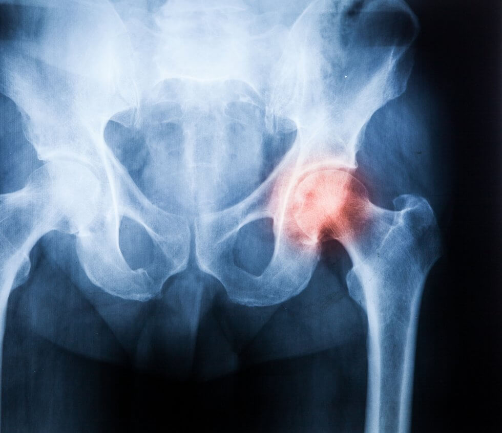
Spinal cord injury is a debilitating, disabling neurological condition resulting from damage to the spinal cord, or to the nerves at the end of the spinal canal. This is most frequently caused by traumatic occurrences such as vehicle accidents, falls, sport injuries or violence, but may also be caused by tumours, infections or degenerative conditions.

Spinal cord injury causes partial or complete loss of sensation and function below the level of the injury, commonly resulting in paralysis (paraplegia or quadriplegia), loss of bladder and bowel control, and breathing issues.[1]

What therapies are currently available?

At present, there are no known therapies that would reverse the initial injury and return an injured spinal cord to full function.[2] Current treatments for spinal cord injury focus on limiting what is called the secondary injury cascade, ideally preventing further damage and thus further loss of feeling and motor function.[3] These methods include surgery, medication, physical therapy and rehabilitation, and assistive devices such as braces or wheelchairs.

What is epidural electrical stimulation for spinal cord injury?

In epidural electrical stimulation, an array of electrodes is implanted along the spinal cord through a surgical procedure called a laminectomy. The stimulation provided by the electrical pulses generated by these electrodes could help in the recovery of functionality following spinal cord injury, improving patients’ ability to walk and stand as well as aiding with bladder and bowel control.[3] [4]

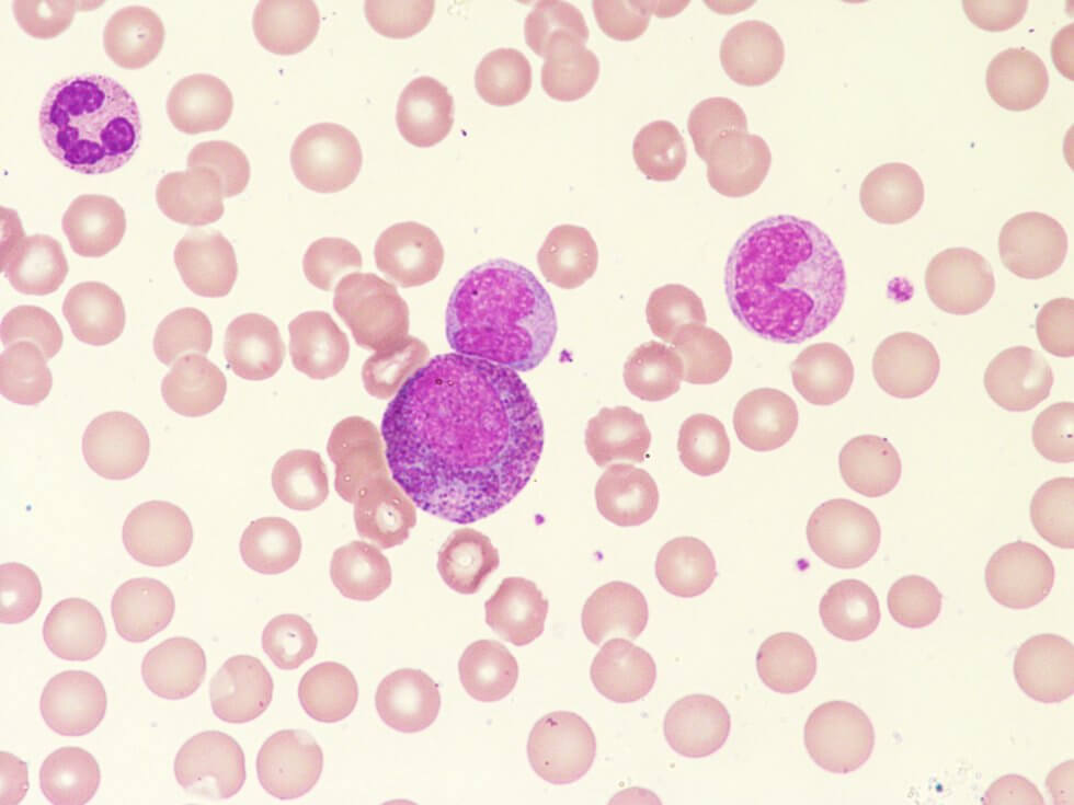
How could stem cells help?

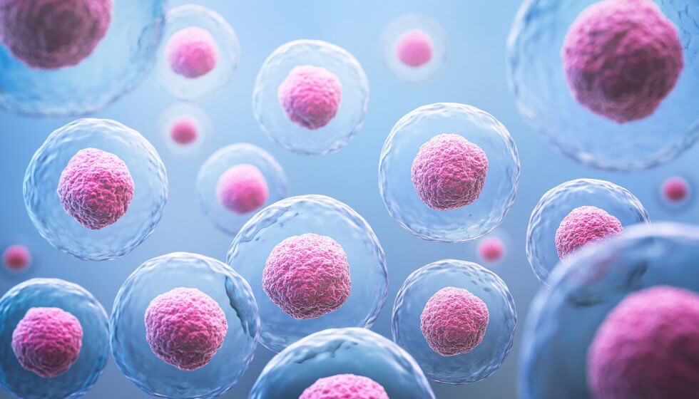
By leveraging the regenerative properties of stem cells, it is hoped that a stem cell treatment could repair and regenerate damaged spinal cord tissue.

This could mean protecting what neurons remain intact, repairing the protective myelin sheath on damaged ones, thus restoring their ability to conduct nerve signals, and replacing lost ones. Stem cells also have the ability to modulate the body’s immune response, and could reduce inflammation and mitigate the secondary damage that follows the initial injury.[5]

There are currently several clinical trials studying the application of stem cells for spinal cord injury. Although more research is needed, results so far are promising, including the high-profile case study of a man who has regained the ability to walk.[6]

What has the new study found?

Researchers at Xi’an Jiaotong University, China, set out to test the effectiveness of a therapy combining both epidural electrical stimulation and stem cell injections, using a mouse model of spinal cord injury.[7]

The study involved four different groups of mice: a group which was treated with EES alone, a group which was treated with a mix of mouse neural stem cells (NSCs) as well as human umbilical cord mesenchymal stem cells (hUCMSCs), a group which received both treatments and a control group in which the spinal cord injury was left untreated. The mice in all groups were monitored and assessed for a period of two months.

Following the injury, all mice had complete loss of function in their hind limbs; a week post-injury, mice in the treatment groups underwent their respective treatments. At the end of the monitoring period, mice in the control group were still unable to support themselves on their hind limbs. Conversely, some mice in the EES group were able to achieve paw standing; mice in the hUCMSC group also achieved this milestone, in a more frequent and sustained manner. Mice in the combined treatment group not only achieved paw standing, but also showed improved motor coordination. Swimming and gait analysis tests corroborated these findings, with the hUCMSC group doing better than the EES group, both doing better than the control and the combined treatment group doing best of all.

What’s next?

Both EES and stem cell transplants are currently the subject of clinical trials to test their effectiveness in the treatment of spinal cord injury. As the new study shows, it is entirely possible that the best treatment will be a combination of both, but more research is required on each individual treatment before the combination therapy can be tested in humans.

What is undeniable is that the number of studies and clinical trials examining the regenerative power of stem cells and their potential for treating currently incurable diseases and injuries continues to grow.

To find out more about what stem cells could do, and how you could preserve a rich source of them for your baby so that they could gain access to future regenerative treatments, fill in the form below to request your free welcome pack.

References

[1] University Hospitals Sussex NHS Foundation Trust. (2023). Spinal cord injury. https://www.uhsussex.nhs.uk/sussex-trauma-network/rehabilitation/conditions/spinal-cord-injury/

[2] NIH (2016). What are the treatments for spinal cord injury (SCI)? https://www.nichd.nih.gov/health/topics/spinalinjury/conditioninfo/treatments

[3] Dorrian, R.M., Berryman, C.F., Lauto, A. and Leonard, A.V. (2023). Electrical stimulation for the treatment of spinal cord injuries: A review of the cellular and molecular mechanisms that drive functional improvements. Frontiers in Cellular Neuroscience, 17. doi:https://doi.org/10.3389/fncel.2023.1095259

[4] Royal National Orthopaedic Hospital. (2024). New research offers quality of life hope for many paralysed after spinal cord injuries. https://www.rnoh.nhs.uk/news/new-research-offers-quality-life-hope-many-paralysed-after-spinal-cord-injuries

[5] Zeng, C.-W. (2023). Advancing Spinal Cord Injury Treatment through Stem Cell Therapy: A Comprehensive Review of Cell Types, Challenges, and Emerging Technologies in Regenerative Medicine. International Journal of Molecular Sciences, 24(18), p.14349. doi:https://doi.org/10.3390/ijms241814349

[6] Stem Cells Help Injured Surfer to Walk Again.

[7] Mu, Z., Qin, J., Zhou, X. et al. (2024.) Synergistic effects of human umbilical cord mesenchymal stem cells/neural stem cells and epidural electrical stimulation on spinal cord injury rehabilitation. Sci Rep 14, 26090. doi:https://doi.org/10.1038/s41598-024-75754-x


hip_pain-980x846-1.jpg

England flanker Tom Curry is undergoing stem cell therapy as a follow-up to last year’s career-saving surgery.[1]

Suffering from chronic hip pain and reduced range of motion, Curry was diagnosed with femoroacetabular impingement syndrome, where the ball of the hip joint is abnormally shaped, causing uneven wear and tear in the joint resulting in cartilage and bone damage.[2][3]

In Curry’s case, there was what was defined as an arthritic change within the hip[4] – labrum and cartilage tears, as well as abnormal bone growth. This was corrected in a six-hour surgery, which reshaped the ball of his hip and repaired the labrum and cartilage via a stem cell transplant.[2]

To fix the tears in Curry’s cartilage, leading hip surgeon Damian Griffin placed a synthetic cartilage graft in the joint and applied stem cells taken from Curry’s bone marrow to it to grow a new surface within the joint.[5] The procedure is performed arthroscopically (keyhole surgery), and it has a much shorter recovery time and carries less risk than surgeries involving metal implants, such as a hip resurfacing, where metal surfaces are used to cover the joint,[6] or a complete hip replacement.[7]

Now, Curry is undergoing another stem cell therapy in a bid to make it to the 2027 Rugby World Cup without needing more surgery.[1] There are fewer details available about this therapy, but it has been stated to be a stem cell injection in his hip to help bone growth.

How can stem cells help with injury recovery? 

Stem cells possess remarkable regenerative potential, which therapies and procedures like those Curry is undergoing take advantage of. The cells that make up bone and cartilage typically derive from mesenchymal stem cells (MSCs), which, in addition to their ability to turn into a variety of specialised cell types, also have anti-inflammatory properties. This makes them ideal to help repair damaged tissue, particularly cartilage, which has very limited regenerative capacity of its own.

Curry is far from the only elite athlete to turn to stem cells to get back to form. Footballer Cristiano Ronaldo used stem cell therapies to successfully address knee problems[8], while tennis great Rafael Nadal treated both chronic knee issues and a long-standing back complaint[8][9]. Boxing legend Mike Tyson also had stem cell therapy.[8]

To find out more about the regenerative power of stem cells, and how your baby’s umbilical cord stem cells could safeguard their health for life, simply fill in the form below to request a free information pack.

References

[1] Jones, C. (2024). Tom Curry: England flanker has stem-cell therapy in bid to make 2027 World Cup. BBC Sport.  https://www.bbc.co.uk/sport/rugby-union/articles/cy47vynl1pvo

[2] Stasko, N. (2024). Tom Curry’s ‘Spectacular’ Comeback After Hip Surgery At HSSH. Harley Street Specialist Hospital.  https://hssh.health/blog/tom-currys-spectacular-comeback-after-hip-surgery-at-hssh/

[3] Aaos.org. (2015). Femoroacetabular Impingement – OrthoInfo – AAOS. https://orthoinfo.aaos.org/en/diseases–conditions/femoroacetabular-impingement

[4] Tom Curry: Sale and England flanker says hip damage ‘a car crash’ but surgery a ‘success’. (2024). BBC Sport. https://www.bbc.co.uk/sport/rugby-union/68288821

[5] Wright, J. (2024). Tom Curry had to have hip ‘stitched back together’ after ‘extensive damage’ done, surgeon reveals. Planet Rugby. https://www.planetrugby.com/news/tom-curry-had-to-have-hip-stitched-back-together-after-extensive-damage-done-surgeon-reveals

[6] Royal Orthopaedic Hospital – Birmingham Hip Resurfacing. https://roh.nhs.uk/services-information/hips/birmingham-hip-resurfacing

[7] NHS (2024). What is a hip replacement? nhs.uk. https://www.nhs.uk/conditions/hip-replacement/what-is-a-hip-replacement/

[8] The Sun. (2020). Tyson, Ronaldo, Nadal and more sports stars who swear by stem cell treatment. https://www.thesun.co.uk/sport/11648461/stem-cell-treatment-tyson-ronaldo/

[9] Associated Press. (2014). Rafael Nadal to have stem cell treatment on injured back, says doctor. The Guardian.  https://www.theguardian.com/sport/2014/nov/10/rafael-nadal-stem-cell-treatment-back


restore_sight-980x653-1.jpg

A recent study published in the Lancet journal has shown that stem cells have been used to bring about significant improvements in vision in what’s being heralded as a world first.

The stem cell treatment focused on the repair of the cornea which becomes damaged in limbal stem-cell deficiency (LSCD), a disease that can lead to blindness.

It’s hoped that these findings could have a huge impact on how those with the disease are treated and that it could underpin future therapy options for those suffering from sight loss. [1]

What is LSCD? 

Limbal stem-cell deficiency (or LSCD) is a disease characterised by the loss or deficiency of stem cells in the limbus, the border between the cornea and the sclera. These stem cells are crucial to the maintenance and repair of the limbus, in addition to ensuring the continuation of its barrier function.

Problems with limbal stem cells can lead to the epithelial breakdown of the cornea resulting in inflammation, scarring and potential vision loss.

LSCD has a variety of causes ranging from genetics to acquired causes like inflammation, infection, and trauma and injury. Management of the disease differs depending on the stage of its progression.

At the early stage, managing symptoms can be sufficient to alleviate its impact on quality of life. However, more progressed instances of LSCD require surgery which usually means transplants from key parts of donor eyes. [2]

What did the stem cell study involve?

The study focused on four patients, two men and two women, aged between 39 and 72 who had all been diagnosed with LCSD in both eyes.

Researchers then derived induced pluripotent stem cells (iPSCs) from donated cord blood and used to fabricate corneal epithelial stem-progenitor cells. These were then cultured and transformed into a thin sheet, iPSC-derived corneal epithelial cell sheets (iCEPS).

After removing a layer of scar tissue covering the cornea in one eye in each of the patients, the iCEPS sheet was transplanted on top and covered with a contact lens to protect the graft.

Patients were then monitored continuously to determine safety outcomes for a period of two years. [3]

What were the results of the stem cell study?

Throughout the whole of the two year safety observation period no serious adverse events occurred. The transplant was accepted by the patients without rejection and without tumour formation. In fact, researchers reported that two of the patients even forwent immunosuppressant drugs.

Following the transplant, all four patients saw immediate improvements in their vision during the first year and three out of four patients experienced sustained improvements in their vision and quality of life beyond one year.

These results are hugely encouraging and the research team, based at Osaka University in Japan, hope to move to a larger scale clinical trial to verify their promising findings. [4]

What does this mean for cord blood banking?

As the results of this trial show, stem cells have huge regenerative potential and are at the forefront of medicine, with researchers still coming to terms with the breadth of their applications.

While this study focused on the use of induced pluripotent stem cells derived from cord blood, other types of stem cells can be found in the umbilical cord and placenta, such as haematopoietic and mesenchymal stem cells, that similarly have enormous potential in the field of regenerative medicine.

Unfortunately, the umbilical cord and placenta are often regarded as mere medical waste, meaning that these stem cells get thrown away. But by storing these stem cells in a process called cord blood banking, you can ensure that your baby always has their own stem cells available for use in future therapies.

If you or someone you know is expecting and wants to know more about the power of stem cells and how they can be stored for future use, fill out the form below to request a free Welcome Pack.

References

[1] Soma, Takeshi et al. (2024). ‘Induced pluripotent stem-cell-derived corneal epithelium for transplant surgery: a single-arm, open-label, first-in-human interventional study in Japan’. The Lancet. doi: 10.1016/S0140-6736(24)01764-1. https://doi.org/10.1016/S0140-6736(24)01764-1

[2] Karakus, S. (2024, August 2). Limbal Stem Cell Deficiency. American Academy of Opthalmology. https://eyewiki.org/Limbal_Stem_Cell_Deficiency

[3] Mallapaty, S. (2024, November 8). World-first stem-cell treatment restores vision in people. Nature. https://www.nature.com/articles/d41586-024-03656-z

[4] Mahindra, A. S. (2024, November 10). Biggest Blindness Breakthrough: Japan Performs World’s First Stem Cell-treatment To Restore Vision. Times Now. https://www.timesnownews.com/health/biggest-blindness-breakthrough-japan-performs-worlds-first-stem-cell-treatment-to-restore-vision-article-115144553


world_ms_day-980x653-1.jpg

The results reported from Phase II of the first FDA-approved trial for the treatment of multiple sclerosis (MS) using stem cells are highly promising, suggesting that stem cells could be a viable treatment option to help improve the lives of those with the condition. [1]

What is the MS trial?

The Phase II trial, which was carried out by The Tisch MS Research Center of New York, constitutes the next stage in the development of the groundbreaking research signalled by the results from Phase I, the first FDA-approved trial to explore the injection of stem cells for MS.

The Phase II trial involved 54 patients split between a test group and a control group in a randomised, double-blind, placebo-controlled study.

Members of each group received 6 injections of either autologous (meaning their own) mesenchymal stem cell-derived neural progenitors (MSC-NPs) or saline placebo every two months.

These injections were administered into the cerebrospinal fluid of multiple sclerosis patients.

The study utilised a cross-over model by which patients who received the stem cell injections in year 1 were then given the saline placebo in year 2 and vice versa.

How does the stem cell treatment work?

Building on their findings from Phase I, researchers identified MSC-NPs for the treatment having ascertained that they promote tissue regeneration and immunomodulatory effects, in addition to being safe and well tolerated. [2]

MSC-NPs are a subpopulation of mesenchymal stem cells that can upregulate growth factors like hepatocyte growth factor (HGF), and minimise ectopic differentiation – essentially, the abnormal differentiation of cells.

By injecting them into the cerebrospinal fluid – a process called intrathecal (IE) injection – researchers found in an experimental autoimmune encephalomyelitis (EAE – an accepted way of modelling the effects of MS) mouse model that MSC-NPs were associated with increased spinal cord myelination, neurological recovery and reduced immune infiltration into the central nervous system. [3]

What were the results?

The results of the Phase II trial demonstrated huge promise for the use of stem cells in treating MS.

Patients requiring walking assistance saw significant improvements in both a timed 25-foot walk test and a 6 minute walking test than those in the control group.

Additionally, treatment recipients demonstrated improved bladder function, with 69% showing improvements in post-void residual volume.

Amongst the other findings were indicators that these stem cells could be helping to restore neuronal cells and reverse cognitive decline in patients with less advanced disease progression.

Furthermore, the stem cell treatment occasioned notable biomarker changes in cerebro-spinal fluid, especially with regards to the decreased levels of CCL2, a protein associated with inflammation, and the increased levels of MMP9, an indicator of the increased presence of reparative cells. [4]

Overall these findings are extremely promising, indicating that stem cells could be used in future treatments that may help to reverse many of the most debilitating aspects of multiple sclerosis, like disability.

The next stage in the study will be for researchers to investigate the effects of increasing the stem cell dosage.

What does this mean for cord blood banking?

This trial is just the latest example of the regenerative potential stem cells have to help treat life-changing conditions like MS.

As exemplified by this study, mesenchymal stem cells in particular, with their immunomodulatory and tissue repair properties, are especially promising.

Incredibly, the umbilical cord and placenta are one of the richest sources of these stem cells, but are routinely thrown away after birth.

By saving these for your baby, you could be giving them the key to future therapies for everything from cancer and stroke to diabetes and heart disease.

The potential offered by stem cells is vast, but you only get one chance to save baby’s stem cells, and that’s the day they’re born.

To find out more about cord blood banking and how it could protect your family’s health, fill out the form below to request a free Welcome Pack.

References

[1] FDA-Approved Phase II Stem Cell Treatment Trial Shows Significant and Diverse Improvements for Multiple Sclerosis (MS) Patients. Tisch MS Research Centre of New York. https://www.tischms.org/phase-ii-analysis-results

[2] Harris, Violaine K. et al. (2018) Phase I Trial of Intrathecal Mesenchymal Stem Cell-derived Neural Progenitors in Progressive Multiple Sclerosis. eBioMedicine, Volume 29, 23 – 30. https://doi.org/10.1016/j.ebiom.2018.02.002

[3] Harris, Violaine K. et al. (2012) Clinical and pathological effects of intrathecal injection of mesenchymal stem cell-derived neural progenitors in an experimental model of multiple sclerosis. Journal of the Neurological Sciences, Volume 313, Issue 1, 167 – 177. https://doi.org/10.1016/j.jns.2011.08.036

[4] Harris, V.K., Stark, J., Williams, A. et al. (2024) Efficacy of intrathecal mesenchymal stem cell-neural progenitor therapy in progressive MS: results from a phase II, randomized, placebo-controlled clinical trial. Stem Cell Res Ther 15, 151 (2024). https://doi.org/10.1186/s13287-024-03765-6


anti_aging-980x426-1.jpg

New research has demonstrated the significant role stem cells play in skin and hair development, which could lead to revolutionary new anti-ageing treatments. [1]

By identifying the ways in which the body grows skin, researchers believe that their findings could lead to the use of artificial skin in treatments for burns, wounds, and age-related degeneration.

The research was conducted as part of The Human Cell Atlas project, which aims to map the genetic instructions contained in every cell in the human body to discover how they work.

New discoveries about skin formation

The study looked at the role immune cells, specifically macrophages, play in the development of skin during pregnancy.

While macrophages – which are a type of white blood cell responsible for killing pathogens – are primarily known for their importance as part of the immune system, researchers sought to identify how they contribute to skin formation in a process known as morphogenesis.

In order to do this, a so called ‘reference atlas’ of prenatal skin (that is, skin that forms between 7-17 weeks after conception) was made using advanced techniques including single cell and spatial transcriptomics – a study of RNA molecules within both one and a population of cells – to detail how elements both within and between skin cells interact.

Their findings showed that interactions between immune cells like macrophages and non-immune cells were crucial for key processes in morphogenesis, including hair follicle development, scarless wound healing, and the formation of blood vessels in the skin. [2]

The important role of stem cells in determining skin formation 

Researchers were able to verify their findings about the significant role played by immune cells in skin morphogenesis by comparing pre-natal and adult skin with a lab-grown skin model derived from stem cells.

The model, known as an organoid, successfully mimicked many aspects of skin and hair follicle development found in prenatal and adult skin.

However, it was only when researchers added macrophages that they found the model created blood vessel networks, confirming that immune cells play a more significant role in the development of skin than previously thought.

This stem-cell derived model of human skin is thought to have a vast array of possible therapeutic applications extending from use in identifying the development of conditions affecting skin and hair to providing a more accurate test material for future skin and hair treatments. [3]

Mesenchymal stem cells – key players in hair and skin formation

Another area researchers looked at during the study was the formation of hair follicles during prenatal development.

In earlier studies with mice, researchers identified special cells called pre-Dc cells that eventually become part of the hair follicle structure (called the dermal condensate, or Dc). [4]

Researchers identified similar cells in human prenatal skin and tracked how they developed into the Dc and another structure called the dermal papilla (Dp), which is important for hair growth.

Using various advanced methods, researchers were able to map out the development of these cells over time and predict their paths.

They found that certain early skin cells (fibroblasts) go down one of two routes: some become specialised for hair follicle formation, while others develop into more general skin cells.

As the hair-specific cells move toward the outer layer of the skin, they turn on genes that help them stick together and form the structures needed for hair growth.

The study also looked at how mesenchymal cells interact with skin’s outer (epithelial) cells during hair follicle formation.

They identified important signalling molecules, such as CXCL12, that help guide cell migration and organisation. Interestingly, immune cells were also predicted to assist with these processes. [5]

Discovering the genetic origins of skin and hair conditions

Having mapped how skin and hair developed in pre-natal skin, researchers turned to investigating the origins of certain skin and hair conditions.

They found that reduced hair growth (hypotrichosis) and abnormal hair shapes (like pili torti) are caused by specific genes that are active during early hair follicle development.

Similarly, they were able to identify that epidermolysis bullosa (EB) – a disorder that makes the skin fragile and prone to blistering – had its roots in the expression of the gene COL7A1 in several types of prenatal skin cells.

Researchers were again able to compare gene expression in prenatal skin with their stem cell-derived lab-grown skin models and found strong similarities, noting that while these diseases often stem from defects in skin structure, they are frequently worsened by immune system involvement. [6]

The importance of stem cells and cord blood banking

As this new research demonstrates, stem cells play a pivotal role in not only helping researchers to better understand the formation of vital organs, like skin, but also in helping to facilitate potential future therapies.

By being able to compare cells from both prenatal and adult skin with artificial lab-grown skin using stem cells, researchers were able to identify the hitherto unknown significance of immune cells in skin formation and accurately map the genetic instructions involved in growing skin.

In their paper published in Nature, The Human Cell Atlas team have achieved something remarkable: essentially providing a set of instructions for creating human skin.

Their research is being heralded as groundbreaking and for good reason. Their findings will help to contribute to the development of a range of therapies for genetic skin conditions, treatments for wounds and burns, and remedies that could help to rejuvenate skin.

While the stem cells utilised in this trial were from embryonic stem cells and induced pluripotent stem cells, the stem cells found in umbilical cord tissue have similarly huge potential in regenerative medicine.

In fact, and as we’ve shown previously, trials utilising mesenchymal stem cells – the same type as found in umbilical cord tissue – are already underway that could aid in the treatment of both hair loss and skin conditions like atopic dermatitis. [7] [8]

What’s clear is that stem cells are pivotal in both cutting edge research and the development of future regenerative therapies.

Unfortunately, the umbilical cord and placenta – both rich sources of stem cells – are routinely thrown away after birth. By saving these powerful stem cells for your baby, you could give them access to future therapies that are currently being developed using stem cell-based research as in this study.

To find out more about saving stem cells for your baby, or the baby of someone you know who’s expecting, fill out the form below to download your free Welcome Pack.

References

[1] Ghosh, P. (2024, October 16). New skin research could help slow signs of ageing. BBC News. https://www.bbc.co.uk/news/articles/c756q166gd9o

[2] Gopee, N.H., Winheim, E., Olabi, B. et al. A prenatal skin atlas reveals immune regulation of human skin morphogenesis. Nature (2024). https://doi.org/10.1038/s41586-024-08002-x

[3] Lee J, Rabbani CC, Gao H, Steinhart MR, Woodruff BM, Pflum ZE, Kim A, Heller S, Liu Y, Shipchandler TZ, Koehler KR. Hair-bearing human skin generated entirely from pluripotent stem cells. Nature. 2020 Jun;582(7812):399-404. doi: 10.1038/s41586-020-2352-3. Epub 2020 Jun 3. PMID: 32494013; PMCID: PMC7593871.

[4] Martisova A, Sommerova L, Krejci A, Selingerova I, Kolarova T, Zavadil Kokas F, Holanek M, Podhorec J, Kazda T, Hrstka R. Identification of AGR2 Gene-Specific Expression Patterns Associated with Epithelial-Mesenchymal Transition. Int J Mol Sci. 2022 Sep 16;23(18):10845. doi: 10.3390/ijms231810845. PMID: 36142758; PMCID: PMC9504245.

[5] Gopee, N.H., Winheim, E., Olabi, B. et al. A prenatal skin atlas reveals immune regulation of human skin morphogenesis. Nature (2024). https://doi.org/10.1038/s41586-024-08002-x

[6] Huitema L, Phillips T, Alexeev V, Igoucheva O. Immunological mechanisms underlying progression of chronic wounds in recessive dystrophic epidermolysis bullosa. Exp Dermatol. 2021 Dec;30(12):1724-1733. doi: 10.1111/exd.14411. Epub 2021 Jun 27. PMID: 34142388; PMCID: PMC9290674.

[7] (2024, January 10). Stem Cells Could Be The Future of Hair Regrowth. Cells4Life. https://cells4life.com/2024/01/stem-cells-could-be-the-future-of-hair-regrowth/

[8] (2024, July 11). Promising Results of Stem Cell Eczema Treatment. Cells4Life. https://cells4life.com/2024/07/promising-results-of-stem-cell-eczema-treatment/


stem_cells-980x560-1.jpg

Recent research indicates that umbilical cord exosomes have huge potential in the field of regenerative medicine.

In this blog we’ll explore what exosomes are, where they’re found, and how they could be used in developing new, life changing therapies for a range of conditions.

What are exosomes?

Exosomes are nano-sized particles that contain key biomolecules like proteins and lipids and travel between cells like messengers, regulating numerous biological processes including inflammatory responses.

Initially thought to be responsible for conveying waste away from cells, in recent years numerous trials and studies have explored their intercellular communicatory function and the  potential this offers in a range of therapeutic applications.

Where are exosomes found?

Exosomes are produced by virtually every cell in the body, including stem cells.

Excitingly they can be derived from the mesenchymal stem cells that exist within both umbilical cord blood and umbilical cord tissue.

Due to the low immunogenicity (the likelihood of being attacked by the immune system when transplanted) of mesenchymal stem cells derived from the umbilical cord, along with their non-invasive harvest procedure and ease of expansion in vitro, umbilical cord MSC exosomes (UC-MSC-Exo) are well suited for research and potentially therapeutic purposes. [1]

What can umbilical cord exosomes do?

Below are a few examples of recent trials utilising UC-MSC-Exo in the treatment of various conditions.

Heal wounds and repair nerves

A 2022 study investigating the use of UC-MSC-Exo in treating cutaneous nerve damage and wound healing showed that exosomes have the ability to promote skin and nerve regeneration.

Studied in vitro, researchers found that UC-MSC-Exo promoted the migration and proliferation of skin fibroblasts – cells that play a key role in forming connective tissue.

Additionally, UC-MSC-Exo were also found to promote the secretion of nerve growth factors by fibroblasts, suggesting that exosomes may enhance wound healing by promoting nerve repair.

Tested in vivo in a mouse model, the study showed that the wounds of the treatment group who received UC-MSC-Exo healed faster than the wounds of the group who received a control medium.

The study concluded that UC-MSC-Exo produced therapeutic effects by promoting skin and nerve regeneration. [2]

Slow the progress of osteoarthritis

Another study from 2022 demonstrated that exosomes derived from the umbilical cord have the potential to be used as a treatment method for osteoarthritis (OA).

Using a rat model, researchers undertook a surgical procedure designed to recreate the effects of knee osteoarthritis. They then administered injections of either UC-MSC-Exo or saline solution. After 8 weeks, knee samples were taken to assess the progression of the disease.

Researchers discovered that in the knees of rats who had received injections of exosomes the progression of OA had been halted and severe damage to knee cartilage prevented.

One of the key reasons behind this, the researchers found, was that the exosomes promoted the migration and proliferation of chondrocytes – cells that make up cartilage. They also found that the exosomes helped to inhibit the secretion of pro-inflammatory factors, as well as regulate immune responses; contributing factors in cartilage regeneration. [3]

Alleviate liver damage and disease

A study from 2020 showed that exosomes derived from umbilical cord mesenchymal stem cells were able to secrete the molecule miR-455-3p which helped to suppress inflammatory immune responses and inhibit signalling pathways in order to improve liver damage. [4]

Another study showed that exosomes derived from umbilical cord mesenchymal stem cells have antioxidant effects.

A contributing factor to the progression of liver damage, oxidative stress occurs when there is an imbalance in free radicals (unstable oxygen molecules) and antioxidants in the liver. Oxidative stress can lead to what’s known as apoptosis – programmed cell death. [5]

By using a mouse model, researchers were able to ascertain that injections of UC-MSC-Exo inhibited apoptosis in liver injury, reversing the fallout from oxidative stress. [6]

Promote heart repair

There have been several studies indicating that exosomes derived from umbilical cord mesenchymal stem cells have the ability to reduce myocardial injury – damage to heart tissue.

A study from 2018 showed that UC-MSC-Exo may have the ability to promote the expression of Smad7, a protein which aids in myocardial protection by blocking certain signalling pathways.

Researchers believed this could be the mechanism underlying exosomes’ ability to inhibit apoptosis, promote angiogenesis (the formation of new blood vessels) and improve cardiac function following acute myocardial infarction. [7] [8]

Rejuvenate older stem cells

An especially intriguing study from 2020 also showed that exosomes derived from umbilical cord mesenchymal stem cells could help in myocardial repair by rejuvenating the activity and function of mesenchymal stem cells in older patients.

Because heart conditions mainly affect older patients, the risk of immunogenicity associated with allogeneic stem cell transplants in helping to treat heart damage is a potential risk. This leaves autologous stem cell transplants – using stem cells from the patient themselves – as an available treatment option. However, due to age and cardiovascular risk factors, these autologous stem cells can be limited in their usefulness.

What this study found, however, is that UC-MSC-Exo have the ability to ameliorate the senescence of older mesenchymal stem cells and renew their biological activity, such as the potential to differentiate into cartilage, bone, and fat cells.

In effect this means that umbilical cord exosomes could help in restoring the regenerative capacity of mesenchymal stem cells in a range of therapies for patients who need them most. [9]

Exosomes and cord blood banking

As some of these trials demonstrate, exosomes are a hugely exciting field of regenerative medicine, and their therapeutic potential is only just beginning to be understood.

Most exciting is that these exosomes can be found in umbilical cord blood and tissue which, although usually thrown away as medical waste, can be stored so that your baby has access to their own stem cells that they can use in future therapies.

To learn more about the potential of umbilical cord stem cells and exosomes, contact our specialist team on 01444 873 950 – they’ll be more than happy to help answer any questions you may have.

In the meantime, why not fill out your details below for a free Welcome Pack containing everything you need to know about storing stem cells for your baby.

References

[1] Zhang, N., Zhu, J., Ma, Q. et al. (2020) Exosomes derived from human umbilical cord MSCs rejuvenate aged MSCs and enhance their functions for myocardial repair. Stem Cell Res Ther 11, 273. https://doi.org/10.1186/s13287-020-01782-9

[2] Zhu, Z., Zhang, X., Hao, H., Xu, H., Shu, J., Hou, Q., & Wang, M. (2022). Exosomes Derived From Umbilical Cord Mesenchymal Stem Cells Treat Cutaneous Nerve Damage and Promote Wound Healing. Frontiers in cellular neuroscience, 16, 913009. https://doi.org/10.3389/fncel.2022.913009

[3] Li, P., Lv, S., Jiang, W., Si, L., Liao, B., Zhao, G., Xu, Z., Wang, L., Zhang, J., Wu, H., Peng, Q., Li, Z., Qi, L., Chi, G., & Li, Y. (2022). Exosomes derived from umbilical cord mesenchymal stem cells protect cartilage and regulate the polarization of macrophages in osteoarthritis. Annals of translational medicine, 10(18), 976. https://doi.org/10.21037/atm-22-3912

[4] Shao, M., Xu, Q., Wu, Z., Chen, Y., Shu, Y., Cao, X., Chen, M., Zhang, B., Zhou, Y., Yao, R., Shi, Y., & Bu, H. (2020). Exosomes derived from human umbilical cord mesenchymal stem cells ameliorate IL-6-induced acute liver injury through miR-455-3p. Stem cell research & therapy, 11(1), 37. https://doi.org/10.1186/s13287-020-1550-0

[5] Li, S., Tan, H. Y., Wang, N., Zhang, Z. J., Lao, L., Wong, C. W., & Feng, Y. (2015). The Role of Oxidative Stress and Antioxidants in Liver Diseases. International journal of molecular sciences, 16(11), 26087–26124. https://doi.org/10.3390/ijms161125942

[6] Jiang, W., Tan, Y., Cai, M., Zhao, T., Mao, F., Zhang, X., Xu, W., Yan, Z., Qian, H., & Yan, Y. (2018). Human Umbilical Cord MSC-Derived Exosomes Suppress the Development of CCl4-Induced Liver Injury through Antioxidant Effect. Stem cells international, 2018, 6079642. https://doi.org/10.1155/2018/6079642

[7] Wang, X., Zhao, Y., Sun, L., Shi, Y., Li, Z., Zhao, X. … Zhu, W. (2018). Exosomes derived from human umbilical cord mesenchymal stem cells improve myocardial repair via upregulation of Smad7. International Journal of Molecular Medicine, 41, 3063-3072. https://doi.org/10.3892/ijmm.2018.3496

[8] Zhao, Yuanyuan, Sun, Xiaoxian, Cao, Wenming, Ma, Jie, Sun, Li, Qian, Hui, Zhu, Wei, Xu, Wenrong. (2015) Exosomes Derived from Human Umbilical Cord Mesenchymal Stem Cells Relieve Acute Myocardial Ischemic Injury, Stem Cells International, 761643, 12 pages, 2015. https://doi.org/10.1155/2015/761643

[9] Zhang, N., Zhu, J., Ma, Q. et al. (2020) Exosomes derived from human umbilical cord MSCs rejuvenate aged MSCs and enhance their functions for myocardial repair. Stem Cell Res Ther 11, 273. https://doi.org/10.1186/s13287-020-01782-9


leukaemia-980x735-1.jpg

Umbilical cord blood has long been recognized as a valuable source of life-saving stem cells.

Since the first cord blood transplant was carried out in 1988 for a patient with Fanconi anaemia, it’s estimated that more than 40,000 cord blood transplants have been delivered worldwide, improving countless lives. [1]

Today, new research is revealing even more potential uses for cord blood, especially in combination with other types of transplants.

One exciting development in this area is the emergence of ‘haplo cord’ transplants, which combine cord blood stem cells with haplo-identical bone marrow, offering promising outcomes and improved survival rates for patients who need stem cell transplants.

What is a haplo cord transplant?

A haplo cord transplant is a procedure that involves the infusion of stem cells from both a cord blood unit and a haplo-identical bone marrow donor.

‘Haplo-identical’ means the donor is a half-match to the patient, typically a parent or sibling.

Both bone marrow and umbilical cord blood contain haematopoietic stem cells (HSCs) which can be used to treat blood cancers like acute myeloid leukaemia (AML) and lymphoma.

HSCs are blood-forming stem cells, meaning that they can differentiate into different kinds of blood cells, including white blood cells, red blood cells, and platelets.

The transplant works by infusing a patient with these blood-forming stem cells so that the patient’s body can begin to produce healthy, rather than diseased, blood cells.

In a haplo cord transplant, the patient is coinfused with both bone marrow stem cells from a related, partially matched donor, along with donated cord blood stem cells.

Haplo cord transplants are not a new concept—they have been under study for over a decade. [2]

In 2014, a Spanish clinical trial demonstrated that the outcomes of haplo cord transplants were comparable to those of traditional matched unrelated donor transplants, putting haplo cord transplants among the frontline for allogeneic stem cell transplant options. [3]

More recently, research led by oncologist Dr. Depei Wu from Soochow University in China has provided more detailed evidence on this transplant approach.

Across trials for AML, B-ALL (B Cell Acute Lymphoblastic Leukaemia) leukaemia and aplastic anaemia it was found that the addition of cord blood to a haploidentical transplant resulted in significant improvements in overall survival. [4] [5] [6]

Why are haplo cord transplants important?

The most exciting and important results demonstrated by studies into haplo cord transplants have been the effect on overall survival rates.

A recent multicentre, phase 3 trial, conducted on patients with acute myeloid leukaemia (AML), showed that those who received a haplo cord transplant had a significantly better three-year overall survival rate compared to those who only received a haploidentical transplant.

Specifically, 80.5% of patients in the haplo cord group survived after three years, compared to 67.8% in the haplo-only group. These results are encouraging for both patients and doctors, suggesting that haplo cord transplants may offer a better chance of long-term survival. [7]

In addition to the improved survival rates, other trials have found that haplo cord transplants also offer other potential advantages.

The addition of cord blood stem cells in transplants has been shown to improve immune responses by accelerating neutrophil (a type of white blood cell that helps the body fight infection) engraftment and platelet recovery. [8] [9]

These outcomes are critical for patients undergoing stem cell transplants, as it means reductions in the risks of infections and complications, including AML relapses.

Additionally, cord blood showed improvements in GvL (graft-versus-leukaemia) outcomes – where donor cells attack leukaemia residing in the host patient – without significantly increasing graft-versus-host-disease (GvHD) rates. [10]

The future of haplo cord transplants

While the early results are promising, there is still much to learn about haplo cord transplants.

Oncologists need to explore various factors, such as the ideal chemotherapy regimen, the role of each donor source, and strategies to minimise complications like graft-versus-host disease.

Additional randomised clinical trials are necessary to better understand the full potential of this transplant approach, however, current research suggests that for patients without a fully matched donor, the combination of cord blood and haplo-identical bone marrow could offer a new hope for survival rates and long-term outcomes.

If you’re interested in learning more about cord blood banking and how it can help in life-saving treatments like haplo cord transplants, fill out the form below to receive a free guide to cord blood banking.

References

[1] Gupta, A. O., & Wagner, J. E. (2020). Umbilical Cord Blood Transplants: Current Status and Evolving Therapies. Frontiers in pediatrics, 8, 570282. https://doi.org/10.3389/fped.2020.570282

[2] (2021, January 28). Haplo-Cord Transplants. Parents’ Guide to Cord Blood Foundation. https://parentsguidecordblood.org/en/news/haplo-cord-transplants

[3] Kwon, Mi et al. (2014, December 1). Haplo-Cord Transplantation Using CD34+ Cells from a Third-Party Donor to Speed Engraftment in High-Risk Patients with Hematologic Disorders. Biology of Blood and Marrow Transplantation. Volume 20, Issue 12. 1083-8791. doi: 10.1016/j.bbmt.2014.08.024

[4] Zhou, B., et al. (2021, January 28). Haploidentical Hematopoietic Cell Transplantation Combined with an Unrelated Cord Blood Unit for Adult Acute Myeloid Leukemia Results in Improved Survival Compared to Haploidentical Hematopoietic Cell Transplantation: Results of a Multicenter, Randomized, Phase III Trial. American Society of Hematology. https://ash.confex.com/ash/2023/webprogram/Paper180993.html

[5] Zhou, B., Xu, M., Lu, S., Liu, Y., Qi, L., Liu, T., Tian, H., Chen, J., Wu, D., & Xu, Y. (2022). Clinical Outcomes of B Cell Acute Lymphoblastic Leukemia Patients Treated with Haploidentical Stem Cells Combined with Umbilical Cord Blood Transplantation. Transplantation and cellular therapy, 28(3), 173.e1–173.e6. https://doi.org/10.1016/j.jtct.2021.12.010

[6] Lei, M., Zhang, Y., Jiao, W., Li, X., Zhou, H., Wang, Q., Qiu, H., Tang, X., Han, Y., Fu, C., Jin, Z., Chen, S., Sun, A., Miao, M., Liu, L., & Wu, D. (2022). Comparison of Haploidentical Hematopoietic Stem Cell Transplant With or Without Unrelated Cord Blood Infusion in Severe Aplastic Anemia: Outcomes of a Multicenter Study. Frontiers in immunology, 13, 912917. https://doi.org/10.3389/fimmu.2022.912917

[7] Zhou, B., Chen, J., Liu, T. et al. (2024). Haploidentical hematopoietic cell transplantation with or without an unrelated cord blood unit for adult acute myeloid leukemia: a multicenter, randomized, open-label, phase 3 trial. Sig Transduct Target Ther 9, 108. https://doi.org/10.1038/s41392-024-01820-5

[8] Kent, Andrew., et al. (2024, August 16). Outcomes of Haplo-Cord Versus Dual Cord Transplants: A Single-Center Retrospective Analysis. Transplantation and Cellular Therapy, Official Publication of the American Society for Transplantation and Cellular Therapy. https://doi.org/10.1016/j.jtct.2024.07.021

[9] van Besien, K., Artz, A., Champlin, R. E., Guarneri, D., Bishop, M. R., Chen, J., Gergis, U., Shore, T., Liu, H., Rondon, G., Mayer, S. A., Srour, S. A., Stock, W., & Ciurea, S. O. (2019). Haploidentical vs haplo-cord transplant in adults under 60 years receiving fludarabine and melphalan conditioning. Blood advances, 3(12), 1858–1867. https://doi.org/10.1182/bloodadvances.2019000200

[10] van Besien, K., Liu, H., Margevicius, S., Fu, P., Artz, A., Chaekal, O. K., … Kwon, M. (2024). Haplo-cord transplant. Realizing the potential of umbilical cord blood grafts. – A review of techniques and analysis of outcomes. Leukemia & Lymphoma, 65(10), 1384–1397. https://doi.org/10.1080/10428194.2024.2361353