Stem Cell Blog

Употребата на матичните клетки од папочна врвца рапидно се зголемува. Пред 10 години крвта од папочна врвца можеше да лекува околу 40 состојби, но денес таа бројка е над 80. Со нетрпение очекуваме нови терапии за болести и нарушувања како што се дијабет, аутизам и мозочен удар, можете да бидете во тек со најновите случувања во регенеративната медицина на нашиот блог за матични клетки.


cell-therapy-for-cerebral-palsy-980x588-1.jpg

A new study on cell therapy for cerebral palsy has, for the first time, directly compared the efficacy of two different treatments. Although more research is needed, the study’s results offer valuable insights for future clinical trials tackling this challenging disorder.

The challenges of cell therapy for cerebral palsy 

Over the past two decades, cell therapy has emerged as a promising treatment for cerebral palsy. In particular, mononuclear cells from cord blood (UCB-MNCs) and mesenchymal stem cells from cord tissue (UCT-MSCs) have proven to be a safe and effective therapy. Many clinical trials have obtained results showing improvements in patients’ motor function through these cells’ anti-inflammatory, neuroprotective and regenerative properties.

However, efforts to pinpoint the best treatment method have been stymied by significant variations in trial parameters. Differences from study to study can include the type and severity of cerebral palsy, the age of the patients, the cell type, dose and delivery method, whether the study is open-label or blinded, and even the methods and time periods used to monitor improvement. Consequently, no two studies are alike, and results cannot be directly compared.[1][2]

Study methods and goal

The new study, conducted in Iran, is a pooled analysis of the results of two studies, one on UCB-MNTs and one on UCT-MSCs. Both studies were conducted in the same research centre; moreover, to aid in the comparison, the research methodology and other variables were kept the same as much as possible.[3][4][5]

The study patients were aged 4-14 and had spastic cerebral palsy with white matter lesions. After eligibility screening, 108 patients were randomly assigned to the two treatment arms or a control group. Treatment was done via intrathecal (into the spinal fluid) injection, with the control group receiving a sham procedure instead. Researchers then assessed patients’ motor function, quality of life, disability and spasticity after 1, 3, 6 and 12 months.

Both individual studies were double-blinded. Furthermore, all statistical analysis, both for individual studies and for the final pooled comparison, was performed by a blinded statistician.

The individual studies aimed to confirm that UCB-MNCs and UCT-MSCs are a safe and effective treatment for cerebral palsy. Following that, the pooled study analysis compared the effects of the UCB-MNC treatment to those of the UCT-MSC treatment across the study period.

Study results

Both the UCB-MNC treatment and the UCT-MSC treatment reported positive results over time when compared to the control group. Patients in both groups showed improvement in gross motor function and quality of life, as well as reduction in disability and spasticity.[4][5]

A graph showing outcomes of cell therapy for cerebral palsy.
Figure 1. A comparison graph of the two different treatment groups and the control group.[3]

As the figure above shows, researchers found that the UCB-MNC treatment group showed stronger improvements in motor function early on. However, both the UCB-MNC and UCT-MSC groups achieved the same level of improvement at 6 months post treatment. At twelve months after treatment, there was some gradual deterioration of the improvements; however, researchers noted that UCT-MSCs did seem to result in more sustainable changes, with patients seeing less deterioration compared to the UCB-MNC group.[3]

Due to this deterioration, researchers posit that repeated doses at regular intervals may be the best route to continued improvement.[1] They also suggest that future trials should investigate treatments combining both UCB-MNCs and UCT-MSCs, as it may prove more effective than individual ones.[3]

The future of medicine

Researchers stress that this study is only a start, and further comparative trials and research are needed. Although both UCB-MNCs and UCT-MSCs offer positive results, there is still no certainty on which cell type and source will prove most effective treatment. This is true not only for cerebral palsy, but also for other illnesses and diseases for which an effective treatment is still being sought.

This uncertainty highlights the importance of comprehensive stem cell banking. By storing as many stem cell sources as possible, you could equip your baby and family with the broadest range of options for future regenerative therapies.

To find out how you could preserve both cord blood and tissue, along with amnion and placenta, for your baby’s potential future use, fill in the form below to request your free guide.

References

[1] Parent’s Guide to Cord Blood. (2025). Cerebral Palsy Response to Cell Therapy as a Function of Time. https://parentsguidecordblood.org/en/news/cerebral-palsy-response-cell-therapy-function-time

[2] Qu, J., et al. (2022). Efficacy and safety of stem cell therapy in cerebral palsy: A systematic review and meta-analysis. Frontiers in Bioengineering and Biotechnology, 10. doi:https://doi.org/10.3389/fbioe.2022.1006845

[3] Nouri, M., et al. (2025). Cell-Based Therapy for Cerebral Palsy: A Puzzle in Progress. PubMed, 26(9), pp.569–574. doi:https://doi.org/10.22074/cellj.2024.2032098.1600

[4] Amanat, M., et al. (2021). Clinical and imaging outcomes after intrathecal injection of umbilical cord tissue mesenchymal stem cells in cerebral palsy: a randomized double-blind sham-controlled clinical trial. Stem Cell Research & Therapy, 12(1). doi:https://doi.org/10.1186/s13287-021-02513-4.

[5] Zarrabi, M., et al. (2022). The safety and efficacy of umbilical cord blood mononuclear cells in individuals with spastic cerebral palsy: a randomized double-blind sham-controlled clinical trial. BMC Neurology, 22(1). doi:https://doi.org/10.1186/s12883-022-02636-y.


amnion_graft_eye_injury-980x653-1.jpg

After an acid attack that “melted” his left eye, Paul Laskey thought he would lose his sight entirely. Now, a series of amnion grafts from a donated placenta has saved his eye, stabilising it and preventing further damage.[1]

A long road to recovery

Mr Laskey, from Newcastle, had acid squirted over his face after confronting a mugger, in February 2023. After he was rushed to hospital, doctors found that the chemical burn had, in effect, “melted” his left eye. The inner and outer layers of his cornea both suffered severe damage; the burn had caused limbal stem cell failure and severe neurotrophic keratopathy. In other words, the eye couldn’t repair or regenerate the cells within the cornea. Furthermore, the eye nerves didn’t work correctly.[2]

Doctors feared Mr Laskey would lose his eye entirely. First, they began by cleaning the eye, removing any remnants of the acid that might have still been there.[3] Then, over the following eight months, Mr Laskey underwent several surgeries, including two cornea transplants.

He also received three amnion grafts to stabilise his eye and help it heal, stopping his sight from deteriorating further. The first graft was a week after the attack, and the last more than a year later, in June 2024.[4]

Amnion grafts and eye injury

Amnion grafts are made from placenta – more specifically, from the amniotic membrane, the thin membrane on the inner side of the placenta which forms the amniotic sac. The amnion has anti-bacterial, anti-inflammatory and anti-scarring properties.[5]

Mothers who deliver via elective C-section at specific hospitals can choose to donate their placenta to NHS Blood & Transplant.[6] Scientists then use these to create specialised tissue grafts – between 50 and 100 from each donated placenta. These grafts are used to treat eye injuries, as well as burns and wounds.[3]

Ophthalmologist prof. Francisco Figueredo, who treated Mr Laskey, said that amnion grafts have been essential in helping the management of his severe eye burn.[3] Having saved his eye and preserved the vision he still has, doctors are now hoping to restore more of his eyesight through stem cell treatment.[1]

The future of placental therapy

Paul Laskey’s story unquestionably highlights the remarkable potential of the placenta and amnion. In addition to treatment for eye injuries, amnion grafts have also helped to save limbs from amputation and aided in facial reconstruction.

Although the regenerative properties of the amniotic membrane have been known for some time, research continues to explore the possible uses of the placenta and its components. Undoubtedly, only time will tell what other potentially life-saving treatments it could be used for.

Amnion patches, for instance, have been used to treat patients with inflammatory heart conditions[7]. Researchers have also used mesenchymal stem cells from the amniotic membrane to 3D print muscles, bone fragments, and even ears[8]. The placenta itself is also rich in mesenchymal stem cells, which are being studied to treat illnesses ranging from Crohn’s disease[9] to osteoarthritis[10].

To learn more about the potential uses of the placenta and other birth tissues, and how you could preserve them for your baby and family’s use in years to come, fill in the form below to receive your free guide.

References

[1] Barbour, S. (2025). Newcastle man’s eye saved with help of placenta after acid attack. BBC News. https://www.bbc.co.uk/news/articles/c8r5zz3yp0go

[2] Harwood-Baynes, M. (2025). Battery acid attack victim reveals how donated placenta treatment saved his eye. Sky News. https://news.sky.com/story/battery-acid-attack-victim-reveals-how-donated-placenta-treatment-saved-his-eye-13312465

[3] Pickover, E. (2025). Donated placenta saves acid attack victim’s eye. The Independent. https://www.independent.co.uk/life-style/health-and-families/acid-attack-eye-placenta-transplant-b2700916.html

[4] Alsford, L. (2025). Man used woman’s placenta to re-build his eye after acid attack. Metro. https://metro.co.uk/2025/02/20/man-used-womans-placenta-re-build-eye-save-sight-acid-attack-22579732/

[5] Cleveland Clinic. Amniotic Membrane: Anatomy, Function & Conditions. https://my.clevelandclinic.org/health/body/amniotic-membrane

[6] NHS Blood and Transplant. (2025). Living amniotic membrane/placenta donation programme. https://www.nhsbt.nhs.uk/what-we-do/transplantation-services/tissue-and-eye-services/tissue-donation/become-a-donor/living-amniotic-membraneplacenta-donation-programme/

[7] Marsh, K.M. et al. (2017). Anti-inflammatory properties of amniotic membrane patch following pericardiectomy for constrictive pericarditis. Journal of Cardiothoracic Surgery, 12(1). doi:https://doi.org/10.1186/s13019-017-0567-7

[8] Kang, H.-W., Lee, S.J., Ko, I.K., Kengla, C., Yoo, J.J. and Atala, A. (2016). A 3D bioprinting system to produce human-scale tissue constructs with structural integrity. Nature Biotechnology, 34(3), pp.312–319. doi:https://doi.org/10.1038/nbt.3413

[9] Jung Pyo Lee, Jung, J., Lee, H., Su Jin Jeong, Kyung Hwa Cho, Seong Ju Hwang and Gi Beom Kim (2012). Comparison of immunomodulatory effects of placenta mesenchymal stem cells with bone marrow and adipose mesenchymal stem cells. 13(2), pp.219–224. doi:https://doi.org/10.1016/j.intimp.2012.03.024

[10] Khalifeh Soltani, et al. (2019). Safety and efficacy of allogenic placental mesenchymal stem cells for treating knee osteoarthritis: a pilot study. Cytotherapy, [online] 21(1), pp.54–63. doi:https://doi.org/10.1016/j.jcyt.2018.11.003


acute_lymphoblastic_leukaemia-980x873-1.jpg

A CAR-T cell therapy for acute lymphoblastic leukaemia[1] has recently been approved by the FDA for use in clinical trials in the United States. The therapy is based on cells derived from allogeneic (donor) umbilical cord blood; it is the very first such treatment to gain FDA approval.[2]

What is acute lymphoblastic leukaemia? 

Acute lymphoblastic leukaemia, or ALL, is a type of blood cancer that starts in the bone marrow. It affects white blood cells (lymphocytes).

In ALL, the bone marrow makes too many of the cells from which lymphocytes derive (lymphoblasts). These lymphoblasts grow and divide quickly; however, they are not fully developed and cannot turn into healthy lymphocytes.

As a result, lymphoblasts build up in the bone marrow and stop all types of healthy blood cells from developing. They may also spread into other parts of the body, including lymph nodes, the liver and spleen, and the brain and spinal cord.[3]

The lack of healthy white blood cells means ALL patients are at a higher risk of infection. ALL can also cause anaemia through the lack of red blood cells, as well as bleeding problems due to lower platelet numbers.[4]

Leukaemia is the most common type of cancer which affects children; of all types of leukaemia, ALL is the most commonly diagnosed in children.[4] It can also affect adults, although this is less common.[3]

What is CAR-T cell therapy?

CAR-T therapy is a type of treatment which uses T cells to fight cancerous cells. The T cells are genetically modified using a viral vector to add a specific protein on their surface, called a chimeric antigen receptor (CAR). This addition makes them able to recognise cancerous cells and destroy them.[5]

In standard CAR-T therapy, the T cells used are the patient’s own, extracted from the blood through a process called apheresis. After extraction, they are taken to a specialised lab to be modified. However, this process is very time-consuming, typically requiring three to five weeks for manufacturing and quality checking. This can be a problem in the case of ALL, as the disease is fast-progressing and could worsen before the CAR-T cells are ready to be reinfused.[6]

What makes this new therapy different?

The new therapy uses T cells derived from allogeneic (someone else’s) cord blood as a starting point instead. These cells are in a more naïve state, meaning they have a higher ability for self-renewal and lower immunogenicity. This means a lower chance to provoke an immune response when used in treatment. As a result, they can be developed into an ‘off-the-shelf’ CAR-T therapy that is ready when needed and does not require a long wait.[2] Following positive results from investigator-initiated trials,[1] the FDA has now approved the therapy for clinical trials in the United States.[2] Furthermore, researchers in China are also testing the therapy in a clinical trial for the treatment of lupus.[7][8]

A similar therapy, also based on T cells derived from donor cord blood, is being developed at the Fondazione Tettamanti research centre in Monza, Italy. Using a special DNA sequence called a transposon, scientists turn the T cells into CAR-enabled cytokine-induced killer cells (CARCIK), making them able to destroy cancer cells. An initial set of clinical trials aimed at determining the feasibility and safety of this treatment showed positive results. Researchers are now continuing to test the therapy in clinical trials for ALL as well as non-Hodgkin lymphoma, and in vitro and animal models for acute myeloid leukaemia.[9]

The benefits of using cord blood for treatments

Being able to derive T cells for CAR treatment from cord blood could pave the way towards more targeted therapies in the future, says Sarah Tettamanti, a researcher at Fondazione Tettamanti. These therapies could be developed from banked cord blood compatible with each specific patient.[9]

For treatments such as these, it could be essential to have ready access to a compatible donor match. Public cord blood banks are available in the UK and worldwide. However, because they rely on donated cords from only a small number of hospitals[10], there is no guarantee that a match will be found when needed.

Conversely, if you store your baby’s cord blood privately at birth, they will always have access to their own perfect match if they ever need it. There is also a 25% chance for the cord blood to be a perfect match for a sibling and 50% chance of a partial match. It is guaranteed to always be a partial match for parents.

It could also prove to be a match for a more distant relative – which thanks to cord blood cells’ naïve state and lower immunogenicity, could still be enough for treatment.

To learn more about the potential uses of cord blood in treatments and how you could preserve this precious resource for your baby and family, fill in the form below and request our free guide.

References

[1] LemanBio (2023). Leman Biotech and Ucello Therapeutics enter into Comprehensive Strategic Collaboration to Advance Universal CAR-T Development. https://www.lemanbio.com/en/news/197.html

[2] BusinessWire. (2025). VectorBuilder Powers FDA IND Approval of World’s First Umbilical Cord Blood-Derived Allogeneic CAR-T Product. https://www.businesswire.com/news/home/20250129313138/en/VectorBuilder-Powers-FDA-IND-Approval-of-World%E2%80%99s-First-Umbilical-Cord-Blood-Derived-Allogeneic-CAR-T-Product

[3] Cancer Research UK (2021). What Is Acute Lymphoblastic Leukaemia (ALL)? https://www.cancerresearchuk.org/about-cancer/acute-lymphoblastic-leukaemia-all/about

[4] Cancer Research UK (2021). About childhood acute lymphoblastic leukaemia (ALL). https://www.cancerresearchuk.org/about-cancer/childrens-cancer/acute-lymphoblastic-leukaemia/about

[5] Cancer Research UK (2021). CAR T-cell therapy. https://www.cancerresearchuk.org/about-cancer/treatment/immunotherapy/types/CAR-T-cell-therapy

[6] Leukemia & Lymphoma Society. Chimeric Antigen Receptor (CAR) T-Cell Therapy. https://www.lls.org/treatment/types-treatment/immunotherapy/chimeric-antigen-receptor-car-t-cell-therapy

[7] https://mp.weixin.qq.com/s/t7tQTUwpl5vmr78cSRXGhQ

[8] https://mp.weixin.qq.com/s/rVx9bVuU3-2pKio_LbO7ZQ

[9] Cerati, F. (2025). Dal cordone ombelicale le cellule modificate per trattare la leucemia. Il Sole 24 Ore. https://www.ilsole24ore.com/art/dal-cordone-ombelicale-cellule-modificate-trattare-leucemia-AGD2eidC

[10] Cord Blood Bank – NHS Blood and Transplant. Where can you donate? https://www.nhsbt.nhs.uk/cord-blood-bank/donate/where-can-you-donate/


stroke_in_the_brain-980x918-1.jpg

A recent study has found that a stem cell therapy could reduce epileptic symptoms after stroke and help the brain recover. The study was performed at the Gladstone Institute of Neurological Disease in California, USA, using a rat model of stroke.
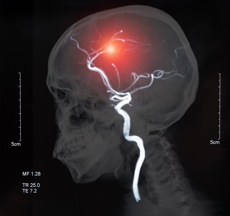
Stroke basics

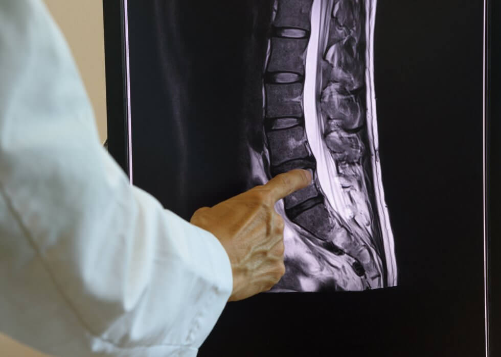
A stroke is a medical condition which causes the death of cells in the brain. This, in turn, stops the brain from working properly. There are two main types of stroke: haemorrhagic, caused by bleeding in the brain, and ischemic, caused by the blockage of a blood vessel. Ischemic stroke is by far the most common, accounting for just under 90% of strokes.[1]

Stroke is the second leading cause of death and one of the major causes of disability worldwide.[2] In the UK, it is the single biggest cause of severe disability, causing a range greater than any other condition, including movement issues, visual problems, and speech difficulties.[3]

The goal of the study

The stem cell therapy tested in the study is based on mesenchymal stem cells derived from donor bone marrow. It has already been successful in clinical trials for improving chronic paralysis after traumatic brain injury[4][5]; this led to its approval as a treatment for this issue in Japan[6].

Now, researchers are hoping it could also help reverse brain damage caused by stroke. In particular, they are focusing on brain hyperexcitability, which is a flawed response in the brain that develops in some people after they have had a stroke.

What is brain hyperexcitability?

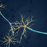
Put simply, brain hyperexcitability is an increased chance for neurons to activate in response to a specific stimulus. In the case of stroke, this flawed response develops in the brain as it tries to make up for lost functions.

These overly active neurons send out signals that are too frequent or too strong to other regions of the brain. This, in turn, causes serious issues. Brain hyperexcitability can make it difficult to control muscles (spasticity) and can lead to seizures.[7] This means stroke is a leading cause of acquired epilepsy,[8] particularly in people over the age of 35.[9]

Unfortunately, brain hyperexcitability remains not well understood, and there is no treatment available to prevent it[7]. Post-stroke seizures and epilepsy are simply managed, as and when needed, with anti-epileptic drugs.[10]

Study process and findings

Researchers tested the stem cell therapy in a rat model of stroke. A month after rats had a stroke, modified human stem cells were injected into their brains, near the damaged area.

The team then measured electrical activity in the brain to determine the effectiveness of the therapy. Furthermore, they also analysed the structure of the brain and blood cells in detail, so they could study the changes wrought by the stroke and by the therapy.

They found that the stroke had caused hyperexcitability in the rats’ brains. The stem cell therapy, however, had reversed this, returning the brain to normal function. Furthermore, a number of proteins and cells that are important for brain function had also increased.

Additionally, by comparing rats which had received the therapy to control rats, researchers identified molecules in the blood that had changed after the stroke. They further saw that these same molecules were restored to normal by the therapy. Additional analysis found that a week after the transplant, few stem cells remained in the rats’ brain; however, the effects were long-lasting.

Hope for future treatment

This therapy is in the very early stages of research; it remains to be determined whether the reversal of brain hyperexcitability would lead to a reduction of symptoms in actual patients.

Still, studies like these highlight the tremendous regenerative potential of stem cells, and the hope they can offer for the treatment of illnesses and injuries that today are considered incurable.

Today, stroke occurs more than 100,000 times per year in the UK – about once every five minutes. Advances in immediate treatment have meant that the number of patients dying from stroke continues to decrease. Unfortunately, however, rehabilitation hasn’t kept pace, and the number of people living with severe disabilities after stroke continues to increase.[3]

Stem cell therapies for post-stroke disability could prove to be truly lifechanging.

Some of the most potent stem cells that could be used in regenerative therapy come your baby’s umbilical cord and placenta. Both of these are normally thrown away at birth – but they could instead be stored for your baby’s future use. If you want to find out more about this rich source of stem cells, and learn how you could preserve it, fill out the form below to download our free parent’s guide.

References

[1] National Heart, Lung and Blood Institute (2023). Stroke – What Is a Stroke? https://www.nhlbi.nih.gov/health/stroke

[2] Katan, M. and Luft, A. (2018). Global Burden of Stroke. Seminars in Neurology, [online] 38(02), pp.208–211. doi:https://doi.org/10.1055/s-0038-1649503

[3] Brain Research UK (2021). Stroke – Neurological condition. https://www.brainresearchuk.org.uk/neurological-conditions/stroke

[4] Kawabori, M. et al. (2021). Cell Therapy for Chronic TBI. Neurology, 96(8), pp.e1202–e1214. doi:https://doi.org/10.1212/wnl.0000000000011450

[5] Okonkwo, D.O. et al. (2024). Mesenchymal Stromal Cell Implants for Chronic Motor Deficits After Traumatic Brain Injury: Post Hoc Analysis of a Randomized Trial. Neurology, [online] 103(7), p.e209797. doi:https://doi.org/10.1212/WNL.0000000000209797

[6] Neuro Central. (2024). SanBio Obtains Marketing Approval for ‘AKUUGO® Suspension for Intracranial Implantation’ (INN: Vandefitemcel) as a Therapeutic Agent for Improving Chronic Motor Paralysis From Traumatic Brain Injury (TBI). https://www.neuro-central.com/sanbio-obtains-marketing-approval-for-akuugo-suspension-for-intracranial-implantation-inn-vandefitemcel-as-a-therapeutic-agent-for-improving-chronic-motor-paralysis-from-tra/

[7] Klein, B. et al. (2024). Modified human mesenchymal stromal/stem cells restore cortical excitability after focal ischemic stroke in rats. Molecular Therapy. doi:https://doi.org/10.1016/j.ymthe.2024.12.006

[8] Adhikari, Y., Ma, C.-G., Chai, Z. and Jin, X. (2023). Preventing development of post-stroke hyperexcitability by optogenetic or pharmacological stimulation of cortical excitatory activity. Neurobiology of Disease, 184, p.106233. doi:https://doi.org/10.1016/j.nbd.2023.106233

[9] Mayo Clinic (2021). Epilepsy – Symptoms and Causes. https://www.mayoclinic.org/diseases-conditions/epilepsy/symptoms-causes/syc-20350093

[10] Holtkamp, M., Beghi, E., Benninger, F., Kälviäinen, R., Rocamora, R. and Christensen, H. (2017). European Stroke Organisation guidelines for the management of post-stroke seizures and epilepsy. European Stroke Journal, 2(2), pp.103–115. doi:https://doi.org/10.1177/2396987317705536


anatomical_heart_model-980x551-1.jpg

Scientists have shown that muscle patches grown from stem cells can be used to strengthen and help repair a failing heart. In a breakthrough clinical trial, ten patches containing 400 million cells were implanted on the heart of a 46-year-old woman who was suffering from heart failure. The results, along with results from earlier studies which tested the same procedure in monkeys, have been published in Nature[1]

What is heart failure?

Heart failure is when the heart is too weak or stiff and, as a result, cannot pump blood around the body as well as it should. It can have a variety of causes, from heart disease and heart attacks to high blood pressure and inflammation.[2][3]

Heart failure is a long-term condition which gradually gets worse over time. Currently, it is considered an incurable condition; it can only be treated to keep the symptoms under control with medication and, sometimes, surgery. In severe cases of heart failure, a heart transplant may be necessary. However, there is a shortage of hearts for transplantation, so patients may have to wait several years before one becomes available.[4]

Development of the therapy

The team of researchers, led by Prof. Zimmermann from University Medical Center Göttingen, Germany, coaxed stem cells to grow into heart muscle and connective tissue cells. They then mixed these cells with collagen gel to create patches which could be applied to the outside surface of the heart using a minimally invasive surgery.

After initial studies in vitro and in small animal models of heart failure confirmed the treatment had potential, the patches were first tested in monkeys. The team implanted the patches into six rhesus macaques with heart failure. Three of the monkeys received two patches, while the other three received five. These monkeys were also all treated with immunosuppressive drugs. A second group of seven monkeys remained untreated as a control.

The implanted cells remained smaller than the monkeys’ own heart muscle cells. However, the patches led to an improvement in heart function compared to the control monkeys, thickening the heart’s muscle and increasing its pumping power.[5]

First in-human trial

The success of the trial in monkeys led to the approval of a first-in-human phase 1/2 trial, called BioVAT-HF, which began in 2021 and is currently ongoing.[6][7][8]

The trial has so far recruited 19 patients. The first of these, a 46-year-old woman, had severe heart failure and was waiting for a heart transplant.[1] The researcher team implanted the muscle patches on her heart; she also received immunosuppressive drugs of the same type normally used for transplants. Three months later, the patient was lucky enough to be the recipient of a successful heart transplant.

Upon analysis of her old heart, scientists found that the implanted patches had survived and had formed blood vessels. In other words, they had integrated with the heart without any side effects.

The therapy is still in the early stages of research, and Zimmermann is very clear that it is not yet a replacement for a heart transplant. Rather, it is a supportive treatment for patients in advanced stages of heart failure, who are waiting for a transplant and are under palliative care.

Still, these are incredibly exciting results, which could prove to be a game-changer for the treatment of heart failure. The research team is continuing the clinical trial. In addition, they are testing new patch designs in monkeys in hopes of minimising the need for immunosuppressive drugs.

Stem cells: the future of medicine

This study is one more example of how stem cells are shaping research through their applications in regenerative medicine.

The stem cells used in this particular clinical trial are allogeneic (donor) induced pluripotent stem cells (iPSCs). When receiving a donor transplant, there is always a risk of rejection, hence the need for immunosuppressive drugs.

Your baby’s umbilical cord is a rich source of stem cells, among the most naïve and potent your baby will ever have. They could be used for similar therapies in the future, should your baby need them. What’s more, they could be used without risk of rejection: they are your baby’s own cells, their own perfect genetic match.

You only get one opportunity to collect and store these cells: in the 15 minutes after your baby is born. To find out more about how you can preserve this precious resource, fill in the form below to request your free guide to cord blood banking.

References

[1] Naddaf, M. (2025). ‘Breakthrough’ stem-cell patches strengthened a woman’s failing heart. Nature. doi:https://doi.org/10.1038/d41586-025-00273-2

[2] NHS (2022). Heart failure. https://www.nhs.uk/conditions/heart-failure/

[3] Mayo Clinic (2023). Heart failure. https://www.mayoclinic.org/diseases-conditions/heart-failure/symptoms-causes/syc-20373142

[4] NHS (2022). Treatment – Heart Failure. https://www.nhs.uk/conditions/heart-failure/treatment/

[5] Jebran, A.-F., et al. (2025). Engineered heart muscle allografts for heart repair in primates and humans. Nature. doi:https://doi.org/10.1038/s41586-024-08463-0

[6] Dzhk.de. (2024). Safety and Efficacy of Induced Pluripotent Stem Cell-derived Engineered Human Myocardium as Biological Ventricular Assist Tissue in Terminal Heart Failure (BioVAT-DZHK20). https://dzhk.de/en/research/clinical-research/dzhk-studies/study/detail/biovathfdzhk20

[7] Gavenis, K., University Medical Center Goettingen, Deutsches Zentrum für Herz-Kreislauf-Forschung (DZHK), University Medical Center Freiburg and Repairon GmbH (2023). Safety and Efficacy of Induced Pluripotent Stem Cell-derived Engineered Human Myocardium as Biological Ventricular Assist Tissue in Terminal Heart Failure. clinicaltrials.gov. https://clinicaltrials.gov/study/NCT04396899

[8] Dzhk.de. (2025). DZHK-Studie BioVAT-HF-DZHK20. https://biovat.dzhk.de


sun_damage-980x842-1.jpg

A recent study, carried out both in vitro and in vivo on a mouse model, has found that extracellular vesicles derived from adipose (fat) and umbilical cord mesenchymal stem cells could have a regenerative effect on sun-damaged skin.[1]

Sun damage causes and consequences

Unprotected exposure to harmful ultraviolet (UV) rays from the sun damages the DNA in your skin, ageing it prematurely. This is called photoaging, or sun damage, and can take several forms:[2] [3] [4]

  • Wrinkles and skin thickening: UV rays break down collagen and elastin, proteins which give your skin its structure and elasticity. As a result, skin becomes thicker and wrinkled over time, beyond what would normally be caused by ageing.

  • Sun spots, redness or blotchiness: skin cells produce melanin as a reaction to UV rays, in an attempt to protect against further sun damage. This is the same process that gives you a tan. However, over time, the damaged skin becomes permanently, unevenly pigmented, causing darker patches often called sun spots. This can also take the form of broken blood vessels, causing permanent redness or blotchiness. This happens particularly in areas frequently exposed to the sun, such as the face and hands.

  • Skin cancer: too much UV exposure can cause skin cancer. In the UK, overexposure to UV radiation is the cause of 85% of cases of melanoma skin cancers.[5]

What are extracellular vesicles? 

Extracellular vesicles are tiny particles which are generated from cells. They can carry important biomolecules, like fats and proteins, to other cells, working as a messenger of sorts.

Because the contents of extracellular vesicles can vary depending on the originating cells, scientists believe they could be of great importance in medicine. They could serve as a prognostic tool to predict the likely course of diseases, as well as help to cure them.[6]

In particular, extracellular vesicles derived from mesenchymal stem cells could inherit their regenerative properties, and have been a recent focus of research for their therapeutic potential.

What did the study find?

In the study, scientists from Peking Union Medical College, Beijing, China, aimed to investigate the effects of extracellular vesicles derived from adipose mesenchymal stem cells (AMSC-EVs) and umbilical cord mesenchymal stem cells (HUMSC-EVs) on photoaging.

In vitro, AMSC-EVs and HUMSC-EVs had positive effects on keratinocytes (cells which make up the outermost layer of the skin) and fibroblasts (cells which make up the connective tissue in the middle layer of the skin) that had been exposed to UV radiation. Treatment with EVs lowered inflammation and reduced the levels at which various biomarkers of senescence (ageing) were present in cells. Moreover, the treatment boosted cell proliferation and migration, properties which make skin cells better able to heal damage. Similar protective and regenerative effects were also observed using an in-vitro, full-thickness model of human skin.

These positive results prompted further analysis in vivo, using nude mice. The mice were randomly split into four groups, keeping one as the control. The other three groups were first exposed to high doses of UV radiation, then treated, respectively, with phosphate-buffered saline (PBS) as a placebo, with AMSC-EVs and with HUMSC-EVs.

All three groups exposed to UV initially developed deep, wide wrinkles. By the end of the observation period, however, the skin of mice treated with EV showed significantly fewer and thinner wrinkles. Skin analysis showed that EV treatment helped the skin recover water content, and reversed the epidermal thickening caused by UV radiation. The treatment also improved collagen and elastin levels and reduced inflammation. Additionally, there were fewer biomarkers of ageing in the skin cells of treated mice.

The benefits of stem cell banking

Both types of EVs studied proved effective in mitigating photoaging. However, the researchers noted that, in the mouse model, the effects of EVs derived from umbilical cord MSCs seemed better, from a therapeutic point of view, than those of EVs derived from fat tissue MSCs. The skin of the mice in the HUMSC-EV treatment group was noticeably less wrinkled. Additionally, the skin’s water content was much closer to that of the mice who had not been exposed to UV radiation at all, as was the epidermal thickness.

Furthermore, the process of collecting fat tissue for therapies is inherently invasive, but must be undergone if patients wish to use autologous (their own) stem cells as a therapeutic source. Conversely, the collection of stem cells from the umbilical cord is a painless, entirely non-invasive process; however, the cord must have been collected immediately after birth, and the cells and tissues cryogenically stored for future use. If this was not done, the only way to access therapies based on umbilical cord stem cells is the use of allogeneic (donor) cords – something which can encounter any number of issues, from lack of availability to incompatibility or rejection.

To find out more about storing your baby’s umbilical cord stem cells, so they will have them ready and waiting rather than needing to seek out alternative stem cell sources should they ever need regenerative therapies, fill in the form below to request your welcome pack.

References

[1] Zhang, H., et al. (2024). Human adipose and umbilical cord mesenchymal stem cell-derived extracellular vesicles mitigate photoaging via TIMP1/Notch1. Signal Transduction and Targeted Therapy, 9(1). doi:https://doi.org/10.1038/s41392-024-01993-z

[2] Grabel, A. (2019). Photoaging: What You Need to Know About the Other Kind of Aging. The Skin Cancer Foundation. https://www.skincancer.org/blog/photoaging-what-you-need-to-know/

[3] Cleveland Clinic (2022). Sun-damaged Skin: Photoaging, Signs, Causes & Treatment.  https://my.clevelandclinic.org/health/diseases/5240-sun-damage-protecting-yourself

[4] Yale Medicine (2023). Photoaging (Sun Damage). https://www.yalemedicine.org/conditions/sun-damage

[5] Cancer Research UK (2020). Risks and causes of melanoma skin cancer. https://www.cancerresearchuk.org/about-cancer/melanoma/risks-causes

[6] Zhang, Y., Liu, Y., Liu, H. and Tang, W.H. (2019). Exosomes: biogenesis, biologic function and clinical potential. Cell & Bioscience, 9(1). doi:https://doi.org/10.1186/s13578-019-0282-2


cord_blood_leukaemia_treatment-980x980-1.jpg

After two planned transplants from unrelated donors fell through at the very last minute, Australian three-year-old Tommy Bacon is now in remission from a rare, dangerous form of leukaemia following a transplant of the stem cells from his baby sister’s cord blood.[1]

Tommy’s story 

Tommy fell ill not long after his parents discovered they were expecting a second child – a baby girl. When he first started showing signs of illness, his parents and their doctor were not immediately alarmed. They assumed it was just a case of the usual germs picked up at daycare, which he had recently begun attending.

After he developed tonsillitis during a family trip to the UK in May 2023, however, his parents took him to the hospital and insisted he should be admitted. Tests eventually revealed that he had leukaemia. More specifically, he had a form of the disease called juvenile myelomonocytic leukaemia (JMML). What’s worse, he had one of the most high-risk, aggressive variants.

JMML is incredibly rare, with only 1-2 children out of one million being diagnosed with it every year[2]. A stem cell transplant is the only curative treatment option. Without a transplant, however, a child with an aggressive variant of JMML could survive for less than a year.[3]

The search for a donor

Australia’s donor registry had no donor compatible with Tommy, so his parents started a donor drive. Eventually, an international donor was found. Unfortunately, however, the donor pulled out of the donation process a week before Tommy was due to start his pre-transplant chemotherapy.

By then, Tommy’s baby sister’s due date was fast approaching, and his parents booked a date for the induction. On the day they were going into hospital, a phone call came that a second donor had been found for Tommy. Still, they decided to have their baby girl’s cord blood collected and stored with an Australian cord blood bank, just in case – although they knew it wasn’t guaranteed that she would be a match for Tommy.

Not long after baby Aria’s birth, the second donor, too, pulled out. The family was heartbroken.

A search for a third donor got underway, but Tommy didn’t have long. Because of this, a decision was made to prepare Tommy’s dad as a half-matched (haploidentical) donor. Such a transplant would not have been ideal, since haploidentical transplant recipients are at higher risk of developing post-transplant complications[4]. Absent a perfect match, though, this was Tommy’s last hope.

Then, the cord blood bank called: they had tested Aria’s cord blood, and she was a perfect match for Tommy.

Within a few weeks, Tommy received his transplant. Four months later, he was in remission.

The importance of family cord blood banking

Tommy’s story highlights the importance of family stem cell banking. By choosing to bank your baby’s cord blood stem cells, they will always be ready and waiting should your baby, or another family member, need them.

Their cord blood stem cells are guaranteed to be their own perfect genetic match. There is also a 25% chance they will be a perfect match for a sibling, and a 50% chance of a partial match. Moreover, they are always a partial match for both parents. This is why it can be so important to bank cord blood for every baby in the family, rather than just one.

Stem cells are being heavily investigated in the field of regenerative medicine to treat a wide variety of illnesses and injuries that are currently considered incurable. There are over 7500 clinical trials currently investigating  both autologous (a patient’s own stem cells) and allogeneic (donor stem cells) uses of stem cells, in the hopes of developing new therapies.

These therapies aim to take advantage of the regenerative qualities of stem cells to aid in healing injuries such as spinal cord damage, heart disease, brain injury, arthritis and type 1 diabetes.

By saving your baby’s cord blood stem cells, you can give your baby and family a better chance of accessing these therapies, should they need one in the future.

“I would strongly recommend that if you’re thinking about getting cord blood collected, do it!” says Tommy’s mum, Kylie. “If it can change a life in such a huge way, why would you not?”[5]

To find out more about how cord blood banking works, and how it could safeguard your family’s health, fill in the form below to request a free welcome pack.

References

[1] Gannon, G. (2025). Aria was a miracle stem cell transplant donor for her brother. The Australian Women’s Weekly. https://www.womensweekly.com.au/news/real-life/stem-cell-transplant-donor/

[2] St. Jude Care & Treatment. Juvenile Myelomonocytic Leukemia Treatment. https://www.stjude.org/care-treatment/treatment/childhood-cancer/leukemia-lymphoma/juvenile-myelomonocytic-leukemia-jmml.html

[3] Lls.org. JMML treatment outcomes. https://www.lls.org/leukemia/juvenile-myelomonocytic-leukemia/treatment/treatment-outcomes

[4] Anthony Nolan. Haploidentical stem cell transplants. https://www.anthonynolan.org/patients-and-families/understanding-stem-cell-transplants/haploidentical-stem-cell-transplants

[5] The Project (2024). Baby Girl’s Stem Cells Save Big Brother’s Life. YouTube. https://www.youtube.com/watch?v=JHwWgqEu_Hs


type_1_diabetes-980x653-1.jpg

A 25-year-old woman from China is the first in the world to have been cured from type 1 diabetes, following an injection of islet cells derived from her own reprogrammed stem cells.

The study, published in the Cell journal, offers real hope to the millions of people worldwide suffering from this disease.

What is type 1 diabetes?

Type 1 diabetes is an autoimmune disease that causes the body’s own immune system to mistakenly attack and destroy insulin-producing cells in the pancreas.[1]

Insulin is a hormone which moves glucose (sugar) from the bloodstream into cells, where it is used for energy, and also helps store any extra glucose.[2]

Without insulin, glucose instead accumulates in the blood, causing a host of severe complications including vision loss, nerve damage, kidney failure, and non-healing ulcers that lead to lower limb amputations.[3]

This means people suffering from type 1 diabetes are dependent on external sources of insulin (through injections or a pump), and have to track and manage their blood sugar levels carefully.

How was the cure developed?

A team at Peking University, Beijing, China, extracted cells from three patients with type 1 diabetes and reverted them to a pluripotent state, from which they could be converted to different cell types in the body. These induced pluripotent stem cells (iPSC) were then used to generate clusters of islet cells, which would then be transplanted back into the patients.

Two and a half months later, the first patient to receive the transplant was producing enough insulin to no longer need external injections; she has remained insulin independent since, for more than a year.

Deng Hongkui, the lead scientist in the study, states the results for the other two patients in the study are also very positive. Those patients would have reached the one-year mark in November; it is hoped that when the results are published the trial could be expanded to more patients.[4]

Why is this trial important?

Islet cell transplantation can be an effective treatment for diabetes. In the past, this has relied on islet cells collected from organ donors, which are not sufficient to meet growing demand and require transplant recipients to use immune-suppressant medication to prevent transplant rejection.

A novel therapy developed by Vertex, currently undergoing clinical trials, aims to solve that issue by deriving islet cells from stem cells; however, this therapy also uses donated stem cells as a source, and thus requires the use of immune-suppressant medication.

It is hoped that the use of autologous (own) stem cells will remove the need for this medication. Since the patient studied was already on immune-suppressants due to a liver transplant, this is not a certainty; because type 1 diabetes is an autoimmune condition, there is still a risk that the immune system could attack the newly-transplanted islets regardless. However, Deng and his team aim to develop islet cells that can entirely evade the immune response.

The importance of banking stem cells

Although further study is required to evaluate the effectiveness of this therapy, breakthroughs like this highlight the potential of stem cells in the development of treatments for life-altering, chronic conditions such as type 1 diabetes.

By banking your baby’s cord stem cells, you can make sure they can be used for regenerative therapies like this, should your baby them in the future. To find out more about saving these powerful cells for your baby, download your free Welcome Pack by filling in the form below.

References

[1] Diabetes UK (2023). What causes type 1 diabetes? https://www.diabetes.org.uk/about-diabetes/type-1-diabetes/causes

[2] Diabetes UK (2022). What is insulin? https://www.diabetes.org.uk/about-diabetes/looking-after-diabetes/treatments/insulin/what-is-insulin

[3] Diabetes UK (2024). Complications of diabetes. https://www.diabetes.org.uk/about-diabetes/complications

[4] Mallapaty, S. (2024). Stem cells reverse woman’s diabetes — a world first. Nature. doi:https://doi.org/10.1038/d41586-024-03129-3


spinal_injury-980x700-2.jpg

After a nick caused by a small shard of glass went unnoticed, Canadian Ron Williams developed a deep, infected wound spanning from the heel to the middle of his foot.

Even after six months of hospital treatment, the wound wouldn’t heal; if the infection had persisted, his leg would have needed to be amputated.

Now, thanks to a pioneering wound-care program involving the application of amniotic membrane from donated placentas, there is barely a scar left[1].

What is the amniotic membrane?

The amniotic membrane, also known as the amnion, is the inner layer of the amniotic sac, which holds the amniotic fluid and protects baby as they grow and develop during pregnancy.

Although it is normally discarded as waste after birth, along with the placenta and the rest of the amniotic sac, the amnion has anti-inflammatory, anti-bacterial and anti-scarring properties[2]; its wound-healing powers were first documented over 100 years ago[1]. It is used in a variety of medical specialisations, including ophthalmology (eye care), wound and burn care, gynaecology and surgery[3]; research suggests it could have properties stimulating tissue regeneration, showing potential for use in regenerative medicine[2].

What does the wound healing treatment entail?

A layer of amnion is applied to the wound, then dressed and left undisturbed; the procedure is repeated once a week until the wound has completely healed. In Mr Williams’ case, the wound healed almost 60% in the first week, and fully closed after five.

Dr Balram Sukhu, director of Mount Sinai Allograft Technologies, which runs the amniotic membrane program at Mount Sinai hospital in Toronto, Canada, says treatment has been provided to more than 80 patients, and has resulted in wound closure in all cases.[1]

Why is this kind of treatment not more widely used?

Dr Mark Jeschke, medical director of the burn program at Hamilton Health Sciences, a hospital network in Canada, says the challenges in bringing this treatment to wider availability lie in the process required to obtain amnion grafts[1]. In Canada, amnion grafts are made from placentas donated by mothers undergoing elective C-section; this is also the case in the UK, where only two hospitals are set up to take donations[4]. While NHS Blood and Transplant provides amniotic membrane grafts for ophthalmic surgery[5], amnion dressings for chronic wound treatment could be harder to come by, and can be pricy, with a NICE document estimating the cost at around £1,000 for a 2x3cm graft[6].

Can you save your placenta?

The alternative to placenta donation is private placenta storage, meaning your placenta, including the amnion, is stored in a private bank so that it can be available for you or your child’s future use, should it be needed.

This is typically done as part of cord blood banking, storing the powerful stem cells from your baby’s cord blood along with cord tissue and the placenta. These cells and tissues are being investigated in numerous clinical trials for their potential in treating a wide variety of injuries and diseases.

If you’d like to find out more about private banking for your baby, fill in the form below to receive your free welcome pack, full of all the details you need to make an informed decision.

References

[1] Alberga, H. (2024). Placenta tissue saved this man’s leg from amputation. How can more people benefit? CTVNews. https://www.ctvnews.ca/health/placenta-tissue-saved-this-man-s-leg-from-amputation-how-can-more-people-benefit-1.7142795

[2] Cleveland Clinic (2024). Amniotic Membrane: Anatomy, Function & Conditions. https://my.clevelandclinic.org/health/body/amniotic-membrane

[3] Munoz-Torres, J.R., et al. (2023). Biological properties and surgical applications of the human amniotic membrane. 10. doi:https://doi.org/10.3389/fbioe.2022.1067480.

[4] NHS Blood and Transplant. (2025). Living amniotic membrane/placenta donation programme. https://www.nhsbt.nhs.uk/what-we-do/transplantation-services/tissue-and-eye-services/tissue-donation/become-a-donor/living-amniotic-membraneplacenta-donation-programme/

[5] Tissue and Eye Services – NHS Blood and Transplant. (2025). Amniotic membrane. https://www.nhsbt.nhs.uk/tissue-and-eye-services/products/eyes/amniotic-membrane/

[6] National Institute for Health and Care Excellence. (2018.) EpiFix for chronic wounds. https://www.nice.org.uk/advice/mib139/chapter/Summary


spinal_injury-980x700-1.jpg

A new study on the effectiveness of treatment for spinal cord injuries indicates that a combination therapy, including epidural electrical stimulation (EES) as well as neural stem cells and umbilical cord stem cells, could offer better results than any one treatment on its own.

What is spinal cord injury?

Spinal cord injury is a debilitating, disabling neurological condition resulting from damage to the spinal cord, or to the nerves at the end of the spinal canal. This is most frequently caused by traumatic occurrences such as vehicle accidents, falls, sport injuries or violence, but may also be caused by tumours, infections or degenerative conditions.

Spinal cord injury causes partial or complete loss of sensation and function below the level of the injury, commonly resulting in paralysis (paraplegia or quadriplegia), loss of bladder and bowel control, and breathing issues.[1]

What therapies are currently available?

At present, there are no known therapies that would reverse the initial injury and return an injured spinal cord to full function.[2] Current treatments for spinal cord injury focus on limiting what is called the secondary injury cascade, ideally preventing further damage and thus further loss of feeling and motor function.[3] These methods include surgery, medication, physical therapy and rehabilitation, and assistive devices such as braces or wheelchairs.

What is epidural electrical stimulation for spinal cord injury?

In epidural electrical stimulation, an array of electrodes is implanted along the spinal cord through a surgical procedure called a laminectomy. The stimulation provided by the electrical pulses generated by these electrodes could help in the recovery of functionality following spinal cord injury, improving patients’ ability to walk and stand as well as aiding with bladder and bowel control.[3] [4]

How could stem cells help?

By leveraging the regenerative properties of stem cells, it is hoped that a stem cell treatment could repair and regenerate damaged spinal cord tissue.

This could mean protecting what neurons remain intact, repairing the protective myelin sheath on damaged ones, thus restoring their ability to conduct nerve signals, and replacing lost ones. Stem cells also have the ability to modulate the body’s immune response, and could reduce inflammation and mitigate the secondary damage that follows the initial injury.[5]

There are currently several clinical trials studying the application of stem cells for spinal cord injury. Although more research is needed, results so far are promising, including the high-profile case study of a man who has regained the ability to walk.[6]

What has the new study found?

Researchers at Xi’an Jiaotong University, China, set out to test the effectiveness of a therapy combining both epidural electrical stimulation and stem cell injections, using a mouse model of spinal cord injury.[7]

The study involved four different groups of mice: a group which was treated with EES alone, a group which was treated with a mix of mouse neural stem cells (NSCs) as well as human umbilical cord mesenchymal stem cells (hUCMSCs), a group which received both treatments and a control group in which the spinal cord injury was left untreated. The mice in all groups were monitored and assessed for a period of two months.

Following the injury, all mice had complete loss of function in their hind limbs; a week post-injury, mice in the treatment groups underwent their respective treatments. At the end of the monitoring period, mice in the control group were still unable to support themselves on their hind limbs. Conversely, some mice in the EES group were able to achieve paw standing; mice in the hUCMSC group also achieved this milestone, in a more frequent and sustained manner. Mice in the combined treatment group not only achieved paw standing, but also showed improved motor coordination. Swimming and gait analysis tests corroborated these findings, with the hUCMSC group doing better than the EES group, both doing better than the control and the combined treatment group doing best of all.

What’s next?

Both EES and stem cell transplants are currently the subject of clinical trials to test their effectiveness in the treatment of spinal cord injury. As the new study shows, it is entirely possible that the best treatment will be a combination of both, but more research is required on each individual treatment before the combination therapy can be tested in humans.

What is undeniable is that the number of studies and clinical trials examining the regenerative power of stem cells and their potential for treating currently incurable diseases and injuries continues to grow.

To find out more about what stem cells could do, and how you could preserve a rich source of them for your baby so that they could gain access to future regenerative treatments, fill in the form below to request your free welcome pack.

References

[1] University Hospitals Sussex NHS Foundation Trust. (2023). Spinal cord injury. https://www.uhsussex.nhs.uk/sussex-trauma-network/rehabilitation/conditions/spinal-cord-injury/

[2] NIH (2016). What are the treatments for spinal cord injury (SCI)? https://www.nichd.nih.gov/health/topics/spinalinjury/conditioninfo/treatments

[3] Dorrian, R.M., Berryman, C.F., Lauto, A. and Leonard, A.V. (2023). Electrical stimulation for the treatment of spinal cord injuries: A review of the cellular and molecular mechanisms that drive functional improvements. Frontiers in Cellular Neuroscience, 17. doi:https://doi.org/10.3389/fncel.2023.1095259

[4] Royal National Orthopaedic Hospital. (2024). New research offers quality of life hope for many paralysed after spinal cord injuries. https://www.rnoh.nhs.uk/news/new-research-offers-quality-life-hope-many-paralysed-after-spinal-cord-injuries

[5] Zeng, C.-W. (2023). Advancing Spinal Cord Injury Treatment through Stem Cell Therapy: A Comprehensive Review of Cell Types, Challenges, and Emerging Technologies in Regenerative Medicine. International Journal of Molecular Sciences, 24(18), p.14349. doi:https://doi.org/10.3390/ijms241814349

[6] Stem Cells Help Injured Surfer to Walk Again.

[7] Mu, Z., Qin, J., Zhou, X. et al. (2024.) Synergistic effects of human umbilical cord mesenchymal stem cells/neural stem cells and epidural electrical stimulation on spinal cord injury rehabilitation. Sci Rep 14, 26090. doi:https://doi.org/10.1038/s41598-024-75754-x