Stem Cell Blog

Употребата на матичните клетки од папочна врвца рапидно се зголемува. Пред 10 години крвта од папочна врвца можеше да лекува околу 40 состојби, но денес таа бројка е над 80. Со нетрпение очекуваме нови терапии за болести и нарушувања како што се дијабет, аутизам и мозочен удар, можете да бидете во тек со најновите случувања во регенеративната медицина на нашиот блог за матични клетки.


stem_cell_treatment_stroke_consequences-980x820-1.jpg

Key details

  • Current treatment limitations: While ischaemic strokes – which account for nearly 90% of all strokes – are currently treated with clot removal and supportive rehabilitation, there remains a significant unmet need for therapies that directly address neurological consequences.

  • Stem cell potential: Mesenchymal stem cells (MSCs), particularly those from the umbilical cord (UC-MSCs), are a key focus in regenerative medicine due to their ability to regulate the immune system, reduce inflammation, and aid tissue regeneration.

  • Treatment development: A recent Phase 2 randomized controlled trial demonstrated that UC-MSC treatment is safe, with patients showing improvements in neurological deficits, quality of life, and functional independence compared to a control group receiving only rehabilitation.

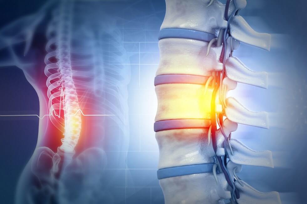
  • Administration route: The trial compared intravenous (IV) and intrathecal (IT) (spinal canal injection) delivery methods, finding that the IV group experienced fewer procedure-related adverse events and demonstrated statistically significant improvements in functional independence earlier (at 6 months) than the IT group.

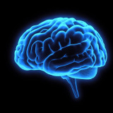
What is an ischaemic stroke?

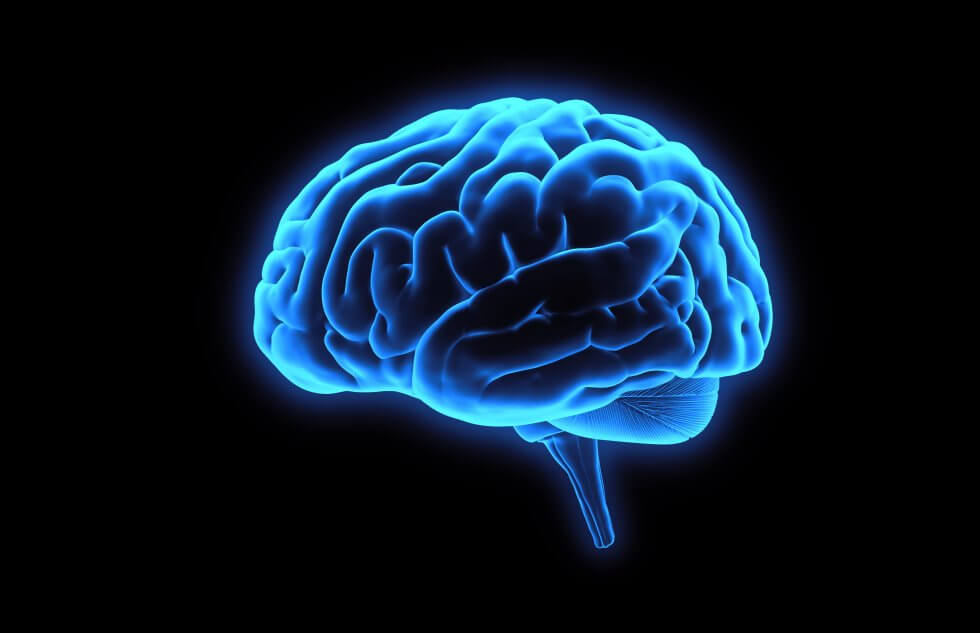
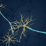
An ischaemic stroke occurs when a clot cuts off blood flow to a part of the brain, preventing brain tissue from receiving oxygen and nutrients. This is a medical emergency, as strokes can happen suddenly and brain cells begin to die within minutes from a blockage happening.[1]

Stroke is a leading cause of death and disability worldwide[2], with ischaemic strokes being the most common, accounting for just under 90% of all strokes.[1]

The consequences of stroke can be severe, and may include:

  • Paralysis

  • Sensory changes

  • Speech or language problems

  • Vision problems

  • Cognitive impairments and memory loss

  • Emotional and behavioural changes.[3]

What treatment is available for stroke and its consequences?

Immediate treatment for ischaemic stroke, provided the patient arrives at the hospital in time, can include:

  • Thrombolysis: using a clot-dissolving medication to break up clots in the brain.

  • Thrombectomy: surgical removal of the clot using a clot retrieval device.[4][5]

This is followed by treatment to reduce the risk of another stroke, such as blood-thinning medication and, potentially, surgery to remedy narrowed arteries.[5]

Treatment for stroke complications is primarily supportive, and focuses on rehabilitation and recovery. Because each stroke is unique, this can vary widely, as it will focus on relearning lost skills and enabling as independent a life as possible. It can involve physiotherapy, speech and language therapy and occupational therapy.[6][7][8]

In other words, recovery and rehabilitation treatment focuses on helping stroke patients adapt to their “new normal”, whatever that may be, leaving a significant unmet need for a treatment that could directly address the neurological consequences of a stroke.

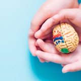
How could stem cells help with the consequences of stroke?

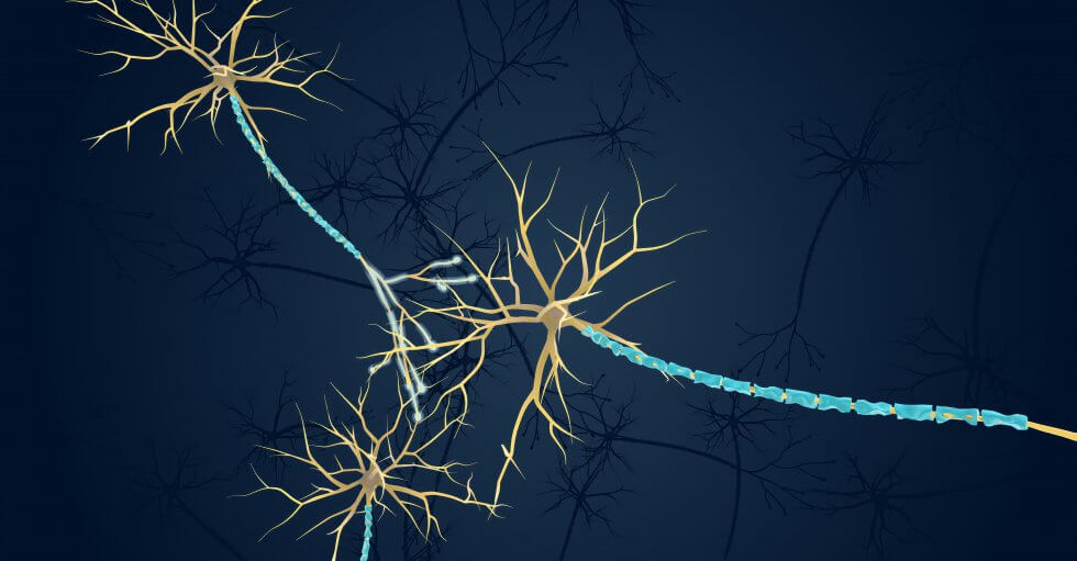
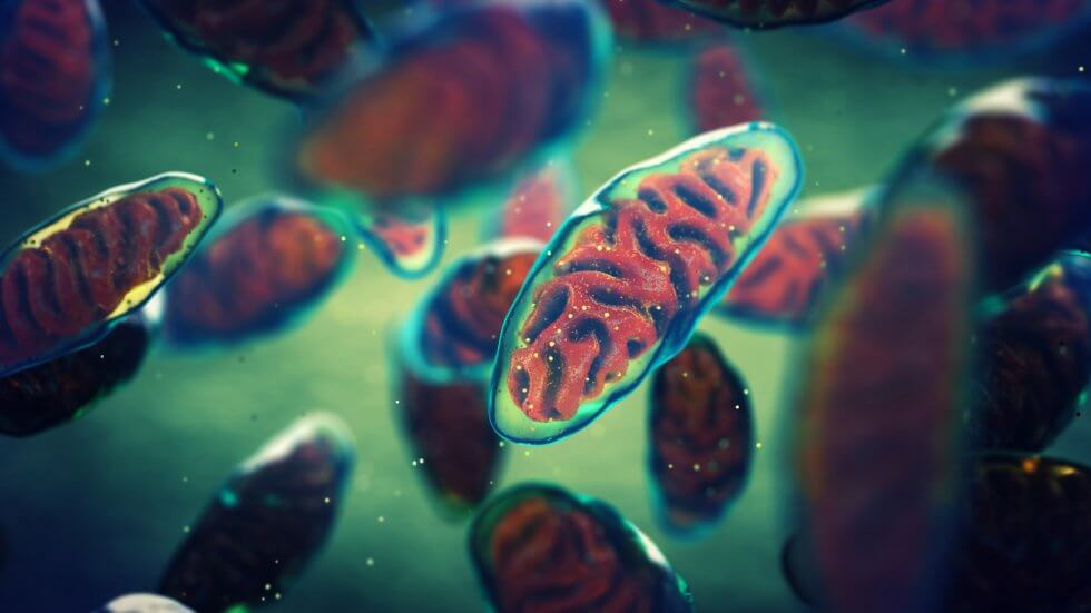
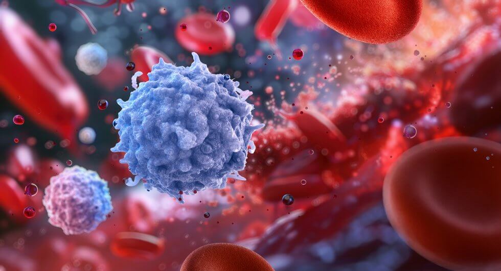
Mesenchymal stem cells (MSCs) are powerful cells which can be obtained from various sources, including bone marrow, adipose (fat) tissue, the umbilical cord and the placenta. These cells have the ability to self-renew and turn into other cell types, such as bone, fat, and cartilage; they can help regulate the body’s immune system and reduce inflammation, promote the formation of new blood vessels, and can release molecules and growth factors which aid in tissue regeneration.[9]

These properties have made them a key focus of the regenerative medicine field, with numerous studies and clinical trials investigating their potential as a treatment for many illnesses and conditions which currently lack effective treatments or cures. MSCs derived from the umbilical cord (UC-MSCs), in particular, have shown in preclinical and clinical studies that they could serve as a safe and potentially effective treatment for the neurological deficits associated with ischemic stroke.[10]

In order to develop a treatment, researchers need to not only identify the best source of stem cells, but also the most appropriate dosage as well as the most effective administration method. It is in this context that a recent clinical trial comparing two different methods of administration places itself.

What were the structure and findings of the clinical trial?

The phase 2 randomised controlled trial evaluated the safety and efficacy of UC-MSCs delivered either via IV (into a vein) or intrathecally (into the spinal canal) for treating neurological deficits after ischemic stroke.

The study involved 32 patients aged 40–75 years who were in the subacute or chronic phase of stroke recovery (7 days to 24 months post-stroke):

  • Patient groups: Patients were randomly assigned to receive UC-MSCs via IV or intrathecally (IT), in addition to standard rehabilitation therapy. A control group consisting of 16 more patients was created by matching patients one-to-one with those in the intrathecal group based on gender, age, and severity of stroke consequences as determined using the NIH stroke scale (NIHSS). This control group only received rehabilitation therapy.

  • Dosage: Patients in the treatment group received a dose of 1.5 × 106 cells per kg of body weight at baseline, and then again 3 months after the first dose.

  • Rehabilitation: Standard rehabilitation therapy consisted of 30 sessions designed to improve cognitive ability, motor function, and overall quality of life, each lasting 60 minutes. The sessions were tailored to each patient and focused on physical, occupational and speech therapy.

  • Monitoring: All patients were followed for a year, with visits scheduled at baseline, 3 months, 6 months and a year.

The results:

  • Safety: There were no severe adverse events related to the treatment. Overall, the adverse event rate was lower in the IV group than in the IT group, where patients experienced more procedure-related pain and headaches.

  • Improvements at 6 months: The IV group demonstrated statistically significant improvements in neurological deficits (NIHSS scores), functional independence, and quality of life compared to the control group. The IT group also showed an improvement compared to the control group, although not as significant.

  • Improvements at 12 months: Both the IV and IT groups showed statistically significant improvements compared to the control group. Researchers hypothesised that IT administration would be more effective than IV, but the study results did not support this assumption – there was no statistically significant difference between the two methods. Larger studies are needed to confirm the results and draw more accurate conclusions.[10]

What are the next steps?

Future studies should focus on optimising the dosage and frequency of the treatment, as well as determining whether any particular patient profile benefits especially well from the treatment. Larger-scale clinical trials will also be necessary to confirm the findings of any smaller-scale trial such as this one.

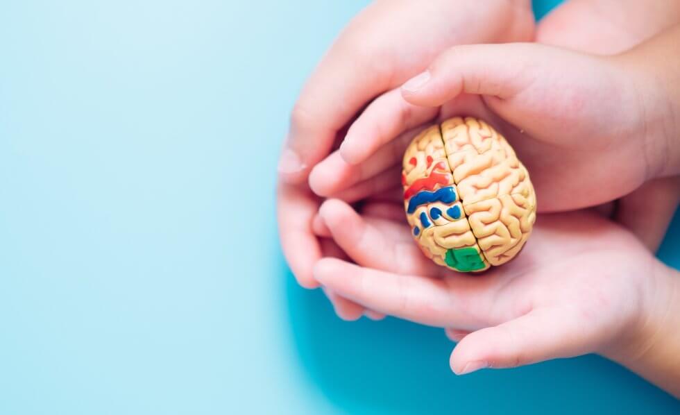
This research, as well as many other such trials investigating stem cell treatments, could come to fruition during your baby’s lifetime. Having access to as many sources of stem cells as possible could be key for them to access such treatments, particularly for conditions such as stroke which can strike very suddenly. To learn more about how you could preserve a rich source of stem cells for your baby as soon as they are born, complete the form below to request our free welcome pack.

References

[1] National Heart, Lung and Blood Institute (2023). What Is a Stroke? https://www.nhlbi.nih.gov/health/stroke

[2] World Health Organization (2021). Mortality and global health estimates. https://www.who.int/data/gho/data/themes/mortality-and-global-health-estimates

[3] American Stroke Association (2025). Effects of stroke. https://www.stroke.org/en/about-stroke/effects-of-stroke

[4] American Stroke Association (2024). Ischemic Strokes (Clots). https://www.stroke.org/en/about-stroke/types-of-stroke/ischemic-stroke-clots

[5] Stroke Association. (2024). Ischaemic stroke treatments. https://www.stroke.org.uk/stroke/types/ischaemic/treatments

[6] American Stroke Association (2024). Post-Stroke rehabilitation. https://www.stroke.org/en/life-after-stroke/stroke-rehab/post-stroke-rehabilitation

[7] Stroke Association (2024). Understanding recovery. https://www.stroke.org.uk/stroke/recovery

[8] American Stroke Association. What to Expect from Stroke Rehabilitation. https://www.stroke.org/-/media/Stroke-Files/Stroke-Resource-Center/Recovery/Patient-Focused/What-to-Expect-from-Stroke-Rehabilitation.pdf

[9] Margiana, R., et al. (2022). Clinical application of mesenchymal stem cell in regenerative medicine: a narrative review. Stem Cell Research & Therapy, 13(1). doi:https://doi.org/10.1186/s13287-022-03054-0

[10] Nguyen, L. T., et al. (2025). Intrathecal versus intravenous umbilical cord mesenchymal stem cells for ischemic stroke sequelae, Stem Cells Translational Medicine, Volume 14, Issue 12, December 2025, zaf063, https://doi.org/10.1093/stcltm/szaf063


stem_cells_charcot_marie_tooth-980x511-1.jpg

Key details

  • New trial approval: The South Korean Ministry of Food and Drug Safety has approved a Phase 2a trial for a new umbilical cord stem cell therapy to treat Charcot-Marie-Tooth disease (CMT), one of the most commonly inherited nerve diseases.[1]

  • Promising phase 1 data: In early trials, patients treated with these stem cells showed significant reductions in disease severity and improvements in gait and balance without serious adverse effects.

  • Orphan drug status: The FDA has granted orphan drug designation to this treatment, highlighting its potential value for rare diseases.

  • Future availability: The developer of the therapy, ENCell, aims for early commercialisation following the completion of the Phase 2 trial.

What is Charcot-Marie-Tooth disease?

Charcot-Marie-Tooth disease (CMT) is a group of inherited disorders that cause damage to the nerves in the arms and legs. It affects both the nerves that control movement (motor nerves) and those that transmit sensations (sensory nerves).[2][3][4]

As one of the most common types of inherited nerve diseases, CMT can be caused by mutations in a wide variety of genes. Depending on the faulty gene, CMT damages either:

  • The myelin sheath, which is the nerves’ protective coat.

  • The axons, which are the nerve fibres themselves.

Both forms of damage significantly impair nerve function.[5][6]

As an inherited condition, CMT runs through families. Most forms of CMT, including CMT1, the main form affecting the myelin sheath and the most common,[6] are inherited in an autosomal dominant pattern. This means someone only needs to get one copy of a faulty gene from one of their parents to develop the condition; inheritance is not connected to the two sex chromosomes, so males and females have equal chances of inheriting the condition. Occasionally, CMT will develop in someone whose family has no prior history, something known as a de novo mutation.[7][8]

CMT is a progressive condition, meaning symptoms can be subtle at first but will get progressively worse over time. Common symptoms include:

  • Mobility issues: difficulty walking, foot drop (trouble lifting the foot at the ankle), and balance issues.

  • Deformities: highly arched or very flat feet, curled toes, and muscle atrophy (shrinking) in the lower legs.

  • Chronic pain: the strain on the body caused by the problems with walking and posture often leads to muscle and joint pain.

  • Hand issues: as CMT advances, it can begin affecting the hands and arms as well, leading to reduced hand dexterity and strength, which can make daily tasks more difficult.[4][9]

There is currently no cure for CMT. Treatment is limited to supportive care and symptom management, including physiotherapy, occupational therapy, orthopaedic devices to support weakened limbs, walking aids such as walkers or wheelchairs, pain medication, and surgery to correct deformities.[3][10][11]

How can umbilical cord stem cells help treat CMT?

The stem cell therapy being trialled for CMT is based on mesenchymal stem cells (MSCs) derived from the Wharton’s jelly found in umbilical cord tissue.[12] Wharton’s jelly is a rich source of powerful MSCs, which, due to their young age, multiply more effectively than MSCs derived from adult tissues such as the bone marrow or fat. They can reduce inflammation, modulate the immune system, and promote healing and regeneration.[13][14]

The therapeutic mechanism involves:

  • Schwann cell support: In laboratory experiments, induced pluripotent stem cells from CMT1 patients were turned into Schwann cells, which are the cells responsible for forming the myelin sheath. Then, the Schwann cells were cultured together with WJ-MSCs, to test whether the latter would have any effect on the former. The results were very positive, showing that WJ-MSCs significantly improve Schwann cells’ ability to multiply.

  • Myelin repair: In mouse models of CMT, mice treated with WJ-MSCs had thicker myelin sheaths and a higher percentage of myelinated neurons compared to control mice.

  • Functional improvements: CMT mice treated with WJ-MSCs also showed improved grip strength and ability to walk.[15]

What do the results of the phase 1 trial for CMT show?

Data from the first-in-human, phase 1 trial of the umbilical cord stem cell therapy for CMT indicates that the treatment is both safe and potentially effective. A total of nine patients received the treatment, three at a low dose and six at a high dose. Results were promising:

  • Safety profile: No serious adverse events, toxicity, or reactions to the treatment were recorded.

  • Symptom reduction: Patients’ score on the Charcot-Marie-Tooth Neuropathy Score version 2 scale, which is used to gauge CMT severity, decreased by an average of 2.89 points after 16 weeks. The high-dose group saw a more marked reduction of 3.50 points.

  • Functional improvements: Patients’ gait, balance, and walking speed also improved.[16]

Following these results, the treatment received an orphan drug designation from the FDA in the in the United States.[17] A phase 1b trial testing multiple doses was already underway when the request for a phase 2 trial was placed and approved;[17][18] the newly combined phase 1b/2a trial aims to further test the treatment against a placebo to confirm its efficacy.[19]

If successful, the treatment would be the first MSC therapy for CMT in the world. ENCell, the company developing the treatment, has said it is aiming for early commercialisation and hopes to bring it to patients as soon as possible after the phase 2 trial is complete.[1] The same therapy is also being investigated for other conditions similar to CMT, including Duchenne muscular dystrophy.[20]

Why is stem cell banking so important?

Stem cell banking ensures that a child has access to their own perfectly matched stem cells, which may be critical for future regenerative therapies.

Much of the research going into stem cells could come to fruition within your baby’s lifetime, providing a cure for illnesses that are currently considered incurable. As development and research are still ongoing, different types and sources of stem cells are being trialled for effectiveness and could turn out to be the key to a specific treatment. This could have an impact on what treatments are available to your baby in the future; the more cell sources and types they have available, the greater their chances of accessing these treatments.

To learn more about stem cells from the umbilical cord and how you could preserve them for your baby’s potential future use, fill in the form below to request your free welcome pack.

References

[1] CHOSUNBIZ (2025). ENCell advances stem cell therapy for Charcot–Marie–Tooth with phase 2a approval. https://biz.chosun.com/en/en-science/2025/11/17/FC2IA63O6NGQTIE6YWPLAQDSW4/

[2] NHS (2019). Charcot-Marie-Tooth disease. https://www.nhs.uk/conditions/charcot-marie-tooth-disease/

[3] Johns Hopkins Medicine. Charcot-Marie-Tooth Disease. https://www.hopkinsmedicine.org/health/conditions-and-diseases/charcotmarietooth-disease

[4] Mayo Clinic (2023). Charcot-Marie-Tooth disease – Symptoms and causes. https://www.mayoclinic.org/diseases-conditions/charcot-marie-tooth-disease/symptoms-causes/syc-20350517

[5] Charcot-Marie-Tooth Association (2025). What Is CMT? https://cmtausa.org/understanding-cmt/what-is-cmt/

[6] NHS (2019). Charcot-Marie-Tooth disease – Causes. https://www.nhs.uk/conditions/charcot-marie-tooth-disease/causes/

[7] CMTUK. (2025). About CMT. https://www.cmt.org.uk/about-cmt/

[8] Charcot-Marie-Tooth Association (2025). Inheritance of Charcot-Marie-Tooth Disease (CMT). https://cmtausa.org/cmt-inheritance/

[9] NHS (2020). Charcot-Marie-Tooth disease – Symptoms. https://www.nhs.uk/conditions/charcot-marie-tooth-disease/symptoms/

[10] NHS (2019). Charcot-Marie-Tooth disease – Treatment. https://www.nhs.uk/conditions/charcot-marie-tooth-disease/treatment/

[11] Cleveland Clinic (2022). Charcot-Marie-Tooth Disease (CMT): Symptoms & Treatment. https://my.clevelandclinic.org/health/diseases/6009-charcot-marie-tooth-disease-cmt

[12] ENCell. Pipeline. https://www.encellinc.com/en/sub/rnd/pipeline.asp

[13] Kim, D.-W., et al. (2013). Wharton’s Jelly-Derived Mesenchymal Stem Cells: Phenotypic Characterization and Optimizing Their Therapeutic Potential for Clinical Applications. International Journal of Molecular Sciences, 14(6), pp.11692–11712. doi:https://doi.org/10.3390/ijms140611692

[14] Drobiova, H., et al. (2023). Wharton’s jelly mesenchymal stem cells: a concise review of their secretome and prospective clinical applications. Frontiers in Cell and Developmental Biology, 11. doi:https://doi.org/10.3389/fcell.2023.1211217

[15] Oh, S.J., et al. (2024). Synergistic effect of Wharton’s jelly-derived mesenchymal stem cells and insulin on Schwann cell proliferation in Charcot-Marie-Tooth disease type 1A treatment. Neurobiology of disease, 203, p.106725. doi:https://doi.org/10.1016/j.nbd.2024.106725

[16] Newswire (2024). ENCell Presents the Phase 1 Clinical Trial Results of EN001 for Charcot-Marie-Tooth Disease at the PNS conference. https://www.newswire.co.kr/newsRead.php?no=992387

[17] Business Wire (2025). EnCell’s EN001 Receives Orphan Drug Designation from the U.S. FDA for Charcot-Marie-Tooth Disease. https://www.businesswire.com/news/home/20250306675394/en/ENCells-EN001-Receives-Orphan-Drug-Designation-from-the-U.S.-FDA-for-Charcot-Marie-Tooth-Disease

[18] Clinicaltrials.gov (2025). Evaluate the Safety and Efficacy of EN001 in Patients With Charcot-Marie-Tooth Disease Type 1A. https://www.clinicaltrials.gov/study/NCT06328712

[19] Kyoung-Won, K. (2025). Korean-made stem cell therapy EN001 enters phase 2a trial for Charcot–Marie–Tooth disease. KBR. https://www.koreabiomed.com/news/articleView.html?idxno=29656

[20] Clinicaltrials.gov (2025). Evaluate the Efficacy and Safety of EN001 in Patients With Duchenne Muscular Dystrophy. https://www.clinicaltrials.gov/study/NCT06328725


umbilical_cord_blood_lesch_nyhan-980x633-1.jpg

Key details

  • Understanding Lesch-Nyhan syndrome: This rare, recessive condition linked to the X chromosome primarily affects boys, and is characterised by a deficiency of the HPRT enzyme, leading to severe motor dysfunction, uric acid overproduction, and compulsive self-injury.

  • Role of HPRT: Used by the body to process and recycle purines: organic compounds that are a type of building block of DNA and RNA.

  • Promising transplant results: A recent case study demonstrated that a cord blood transplant (from a 5/6 HLA-matched donor) successfully restored HPRT1 protein levels to normal range and prevented the onset of self-injury behaviour.

  • Cord blood efficacy: Umbilical cord blood is often considered the preferred stem cell source for treating inborn errors of metabolism because it supports successful engraftment and is safer than other sources when the donor and patient are not a perfect genetic match.

What is Lesch-Nyhan syndrome?

Lesch-Nyhan syndrome is a rare condition which occurs almost exclusively in boys. It is classified as an inborn error of metabolism, meaning it is congenital (present at birth), inherited, and involves a malfunction in the body’s chemical processes (metabolism). It affects a child’s brain and behaviour, as well as causing overproduction of uric acid, a waste product which is normally eliminated through the kidneys.[1][2][3][4] Symptoms include:

  • Urate crystals: Forming in the urine due to the build-up of uric acid, these orange-coloured crystals can deposit in the diapers of babies with this condition. They are often the first symptom to appear.

  • Motor issues: These begin to manifest around four months of age, with decreased muscle tone (hypotonia) resulting in a limp, “floppy” appearance and poor head control being one of the earliest symptoms. This is followed by involuntary muscle spasms (dystonia), repetitive movements (chorea) and flailing of the limbs (ballismus), as well as muscle rigidity (spasticity). Opisthotonos, a severe muscle spasm which causes the back to arch and the head and heels to bend backwards, can also occur. Babies can miss developmental milestones such as sitting, crawling or walking; those who had previously learned to sit upright typically regress and lose the ability. Most people with Lesch-Nyhan syndrome cannot walk, are unable to sit without support, and generally use a wheelchair. Speech can be slurred or poorly articulated (dysarthria); problems with swallowing (dysphagia) can also occur.

  • Gout: The accumulation of uric acid in the joints eventually leads to recurring pain and swelling, similar to what happens in adults with gout. These episodes generally begin in the late teens to early adulthood, and can become progressively more frequent over time.

  • Intellectual disability: Some cognitive impairment is typically present, although it is hard to gauge accurately due to the motor issues and dysarthria.

  • Self injury: This compulsive behaviour is a hallmark of Lesch-Nyhan syndrome, occurring in a majority of patients (about 85%), and typically begins in early childhood. It includes biting of the cheeks, lips, fingers or hands, as well as banging the head or limbs against hard objects, scratching the face, or poking at eyes.

  • Behavioural issues: Patients with Lesch-Nyhan syndrome may also have episodes in which they act aggressive and attempt to injure others, or become verbally abusive.[3][4]

What causes Lesch-Nyhan syndrome?

Lesch-Nyhan syndrome is caused by a genetic mutation, and is inherited in an X-linked recessive pattern. The affected gene, HPRT1, is located on the X chromosome; this means that females are mostly carriers as they’re often protected by an unaffected X chromosome, and males who inherit the defective chromosome from their mothers are affected by the condition. Occasionally, Lesch-Nyhan syndrome can also develop in a family with no history of it. This is known as a spontaneous, or de novo, mutation.

Whether inherited or spontaneous, the mutation results in a severe shortage or complete absence of the HPRT1 protein. The body uses this protein to process and recycle purines, organic compounds which are a type of building block of DNA and RNA. Without this protein, purines are instead broken down but not recycled, resulting in abnormally high levels of uric acid in the blood. When too much uric acid is produced, the kidneys can’t keep up, and uric acid accumulates in the body.[1][2][3][4]

It is still unclear how Lesch-Nyhan syndrome affects the brain, causing the neurological and behavioural issues associated with the condition. Researchers suspect that it may either impact the levels of dopamine, an important chemical messenger required for the brain to function correctly, or reduce the function of dopamine receptors.[2][4]

Is there a cure for Lesch-Nyhan syndrome? What treatment is available?

There is no cure for Lesch-Nyhan syndrome. Treatment primarily aims to treat symptoms, and is generally handled by a team of specialists rather than a single treating physician.[2][3]

Treatment can include:

  • Allopurinol, a medication normally used for gout, to reduce the amounts of uric acid present in the body and control the symptoms caused by it.

  • Procedures to break down kidney or bladder stones.

  • Physical splints or restraints to prevent self-injury, including hip, chest and elbow restraints as well as a mouth guard.

  • Supportive equipment such as a wheelchair.[2][3]

No standard treatment is available for the neurological and behavioural symptoms of Lesch-Nyhan syndrome, although a variety of medication may help ameliorate some of them.[2][3][5][6]

Could a cord blood transplant help with Lesch-Nyhan syndrome?

If performed early enough, a cord blood transplant could potentially slow or halt the progression of Lesch-Nyhan syndrome. A recently published case study, detailing the results of a cord blood transplant for Lesch-Nyhan syndrome, provides the strongest evidence to date for this.[7]

The patient, a boy, initially presented at six months of age with hypotonia, inability to roll over, and occasional opisthotonos. MRI and EEG results raised the suspicion of Lesch-Nyhan syndrome, which was confirmed with genetic testing. Crucially, the diagnostic confirmation happened before the onset of any self-injury compulsion.

As the condition was progressing and the patient did not have a matched sibling donor, doctors made the decision to proceed with a cord blood transplant from a partially matched (5/6 HLA) donor, performed at 14 months old.

The outcome of the transplant was positive:

  • Treatment safety: 28 days post-transplant, the patient had mild graft-versus-host disease (GvHD), a condition in which white blood cells remaining in the donated graft attack the cells of the host’s body. This was resolved with corticosteroids, a medication which reduces inflammation and suppresses the immune system. Beyond this, there were no complications, and the boy remained free of GvHD by the time the case report was submitted for publication.

  • Successful engraftment: Complete donor chimerism, meaning over 95% of the patient’s new blood cells were of donor origin, was achieved on day 32 post-transplant.

  • HPRT1 protein levels: Prior to the transplant, the patient’s HPRT1 protein levels were low (40.9 pg/ml) compared to those of his father and mother (98.8 pg/ml and 78.9 pg/ml respectively). Post-transplant, levels rose to 91.1 pg/ml by day 32 and remained normal (97.0 pg/ml) at day 126.

  • Neurological improvement: The patient’s dystonia and spasticity decreased, opisthotonos became more infrequent, and he had improved facial expressions, increased social interaction and developmental progress. At 36 months of age, he still showed no sign of any self-injury behaviour.[7]

This case shows a promising result, but research is still in the very early stages. Including this case, only three haematopoietic stem cell transplants (HSCT) have been performed in children with Lesch-Nyhan syndrome before the onset of the self-injury compulsion:

Endres et al. (1991) Kállay et al. (2012) Weng et al. (2025)
Patient age 16 months 24 months 14 months
Stem cell source Bone marrow Cord blood (6/6 match) Cord blood (5/6 match)
Outcome Death (day +10) Survival, full chimerism Survival, full chimerism
Long-term outcome N/A No self-injury at 5yr follow-up. Persistent motor delay. No self-injury at 36mo. Improved dystonia/social.

Thus, no formal protocol for this treatment has been established yet. Instead, until more evidence is available, the decision on whether a transplant is appropriate should be made individually for each case, by a coordinated multidisciplinary team including ethical oversight, with transparent family counselling so that informed parental consent is possible.[7]

What is known for certain is:

  • HSCT has been successfully used to treat other conditions in the inborn error of metabolism category, and is the standard of care for some of them, such as Hurler syndrome [8] and Krabbe disease.[9]

  • Cord blood is generally the preferred stem cell source for such transplants, as it increases the likelihood of full donor chimerism and is safer to use when the donor and the patient aren’t a perfect genetic match.[7][8][10]

In the case of Krabbe disease, specifically, early transplantation is of paramount importance; the sooner a transplant happens, the better the functional results for the child, with differences being noticeable even between children transplanted before 30 days of age and those transplanted after.[11] In the United States, several states have implemented newborn screening for Krabbe disease because of this. Here, too, cord blood presents itself as a superior stem cell source, as families may have previously banked the cord blood from an unaffected sibling and, if not, a matched cord blood unit may be available in a public bank.

Should a newborn screening targeted at Lesch-Nyhan syndrome be implemented, it would mean the condition could be identified before symptoms develop. The authors of this case study speculate that early transplant could prevent irreversible neuronal loss and preserve neurological potential.[7]

The importance of family banking

Storing your baby’s cord blood privately gives them access to their own perfectly matched stem cells, and a 75% chance they could be a match for a sibling who may need a transplant; comprising a 25% chance of a perfect match, and 50% chance of a partial match.

As the regenerative medicine field continues to advance, researchers are also investigating the potential of autologous stem cell treatments, using patients’ own stem cells. These would involve gene editing techniques such as CRISPR, and have already become available for some inherited conditions, such as sickle cell disease and beta thalassemia; for other conditions, including Lesch-Nyhan[12] and Krabbe disease, they are in pre-clinical or clinical trial stage. For any of these conditions, it is also possible that the most effective treatment would be a combination of cord blood transplant and gene therapy; this is currently being investigated for Krabbe disease.[13]

Should you choose to bank your baby’s cord blood, it is possible that the stem cells it contains could be used for autologous treatments, rather than more invasively collected bone marrow or peripheral blood stem cells. Should your baby prove to be a donor match for one of their siblings or another family member who is in need of a transplant, the cord blood could potentially also be used in that case.

To learn more about banking your baby’s cord blood, as well as about other sources of stem cells that can only be collected immediately after birth, fill in the form below to request your free guide.

References

[1] MedlinePlus. Lesch-Nyhan syndrome. https://medlineplus.gov/genetics/condition/lesch-nyhan-syndrome/

[2] Cleveland Clinic (2022). Lesch-Nyhan Syndrome: Causes, Symptoms & Treatment. https://my.clevelandclinic.org/health/diseases/23493-lesch-nyhan-syndrome

[3] National Organization for Rare Disorders (2015). Lesch Nyhan Syndrome. https://rarediseases.org/rare-diseases/lesch-nyhan-syndrome/

[4] Nanagiri, A. and Shabbir, N. (2020). Lesch Nyhan Syndrome. https://www.ncbi.nlm.nih.gov/books/NBK556079/

[5] Brainfacts.org. (2025). Lesch Nyhan Syndrome. https://www.brainfacts.org/diseases-and-disorders/neurological-disorders-az/diseases-a-to-z-from-ninds/lesch-nyhan-syndrome

[6] Nyhan, W.L. (2005). LESCH-Nyhan Disease. Journal of the History of the Neurosciences, 14(1), pp.1–10. doi:https://doi.org/10.1080/096470490512490

[7] Weng, T.-F., Tin, C.-H. and Wu, K.-H. (2025). Umbilical Cord Blood Transplantation in Lesch-Nyhan Syndrome: A Case Report and Literature Review. Cureus. doi:https://doi.org/10.7759/cureus.97008

[8] Tan, E.Y., Boelens, J.J., Jones, S.A. and Wynn, R.F. (2019). Hematopoietic Stem Cell Transplantation in Inborn Errors of Metabolism. Frontiers in Pediatrics, 7. doi:https://doi.org/10.3389/fped.2019.00433

[9] Wright, M.D., Poe, M.D., DeRenzo, A., Haldal, S. and Escolar, M.L. (2017). Developmental outcomes of cord blood transplantation for Krabbe disease. Neurology, 89(13), pp.1365–1372. doi:https://doi.org/10.1212/wnl.0000000000004418

[10] Aldenhoven, M. and Kurtzberg, J. (2015). Cord blood is the optimal graft source for the treatment of pediatric patients with lysosomal storage diseases: clinical outcomes and future directions. Cytotherapy, 17(6), pp.765–774. doi:https://doi.org/10.1016/j.jcyt.2015.03.609

[11] Allewelt, H., Taskindoust, M., Troy, J., Page, K., Wood, S., Parikh, S., Prasad, V.K. and Kurtzberg, J. (2018). Long-Term Functional Outcomes after Hematopoietic Stem Cell Transplant for Early Infantile Krabbe Disease. Biology of Blood and Marrow Transplantation, 24(11), pp.2233–2238. doi:https://doi.org/10.1016/j.bbmt.2018.06.020

[12] Jang, G., Shin, H.R., Do, H.-S., Kweon, J., Hwang, S., Kim, S., Heo, S.H., Kim, Y. and Lee, B.H. (2023). Therapeutic gene correction for Lesch-Nyhan syndrome using CRISPR-mediated base and prime editing. Molecular Therapy – Nucleic Acids, 31, pp.586–595. doi:https://doi.org/10.1016/j.omtn.2023.02.009

[13] ClinicalTrials.gov (2024). A Phase 1/2 Clinical Study of Intravenous Gene Transfer With an AAVrh10 Vector Expressing GALC in Krabbe Subjects Receiving Hematopoietic Stem Cell Transplantation (RESKUE). https://www.clinicaltrials.gov/study/NCT04693598


world_cord_blood_day_2025-980x653-1.jpg

Today is World Cord Blood Day! A lot of progress has been made since the very first cord blood transplant was performed in 1988, 37 years ago. Since then, over 60,000 cord blood transplants have been performed worldwide, and cord blood stem cells have become the accepted treatment for over 80 illnesses and conditions, ranging from Fanconi anaemia to Krabbe disease, from SCID to leukaemia, and more.

What’s more, thousands of clinical trials all over the world are investigating the potential of cord blood stem cells to treat conditions which are currently incurable, including cerebral palsy, heart failure, spinal cord injuries and Crohn’s disease, to name but a few.

Despite this, cord blood and other perinatal sources of powerful stem cells, including the cord tissue and the placenta, are still frequently discarded after birth. Thus, World Cord Blood Day was created to highlight cord blood’s therapeutic potential and encourage more families to preserve it for the future.

Cord blood news

Research in the regenerative medicine field continues to advance our knowledge of what’s possible using cord blood. In case you’ve missed them, here are some news articles from the past year:

Banking your baby’s cord blood

Both public and private banks are an option if you want to save your baby’s cord blood rather than having it discarded.

If you choose a public bank, you are donating your baby’s cord blood. This means that it will be available for use to anyone who might need it. The NHS accepts cord blood donations if you are giving birth at one of three hospitals; the Anthony Nolan charity can collect cord blood donations from five more.

As an alternative, you can choose private cord blood banking. When you do this, your baby’s cord blood stem cells will be stored and reserved solely for your family’s use, ready and waiting should your baby, or another family member, ever need them. In addition to being a perfect genetic match for your baby, they also have a 25% chance of being a perfect match for a sibling, and a 50% chance of being a partial match. They are also guaranteed to be a partial match for parents.

At Cells4Life, we offer a range of services aimed at providing the maximum possible benefit to families who choose to store their baby’s precious stem cells with us. Our proprietary processing technology retains up to 3 times more stem cells than industry-standard processing methods, making cord blood collection fully compatible with delayed and optimal cord clamping. We also offer cord tissueplacenta and amnion banking.


stem-cell-treatment-brain-damage-newborns-980x598-1.jpg

A recent study conducted at UMC Utrecht, Netherlands, has shown that mesenchymal stem cells (MSCs) are a safe treatment for brain damage caused by stroke in newborns, and could have a significant positive effect.

Perinatal stroke and its consequences

Perinatal stroke is defined as a stroke that happens to unborn or newborn babies before, during, or shortly after birth, typically between the last few months of pregnancy and one month of age.[1] It is estimated to affect about 1 in 1000 to 3000 children at birth[1][2], although the real figure may be higher as it is likely underdiagnosed.[2]

Perinatal stroke, particularly perinatal arterial ischemic stroke (PAIS) which is the most common type (70% of cases)[2], can have serious consequences. Depending on the artery involved, 50-70% of children who suffered a perinatal stroke will have lifelong issues, including cerebral palsy, epilepsy, vision issues, cognitive impairment and language delays.[3] There is currently no treatment available that can alleviate these issues; the current standard of care is limited to supportive measures.[2][3] Thus, there is an urgent need to develop an effective regenerative treatment and ensure children who suffered a stroke get the best possible start in life.

The PASSIoN study

A research team at UMC Utrecht has been focusing on developing a treatment for perinatal stroke using MSCs.[3] These powerful stem cells offer several advantages, boasting neuroregenerative and anti-inflammatory properties that could prove key to reducing the impact of stroke on a newborn’s brain.[4] Administration of the MSCs via nose drops was selected as being the method of treatment that could potentially deliver the most stem cells to the brain.[3][4]

Following on from preclinical studies in mice, researchers set up a phase 1 clinical trial, called PASSIoN (Perinatal Arterial Stroke treated with Stromal cells IntraNasally), to determine the safety of the treatment.

Newborns were eligible for inclusion into the trial if they were born at any of the neonatal intensive care units in the Netherlands and showed symptoms of PAIS, including seizures and breathing difficulties. Eligible newborns were transferred to UMC Utrecht, where an MRI was performed to confirm whether they had a stroke; if so, they received the stem cell treatment within 7 days after first showing symptoms. In total, ten newborns received the treatment between February 2020 and April 2021.[3]

What were the results?

Initial results for the study were published in 2022, in the prestigious The Lancet journal. The treatment proved to be safe, with no serious adverse events found either immediately after treatment or at the three-month follow-up visit. One newborn did develop a high fever after treatment, but it passed spontaneously within a few hours. Moreover, an MRI scan done at the three-month follow-up showed no unexpected brain abnormalities.

More recently, researchers published long-term follow-up results for the study, along with a comparison between the study patients and a cohort of newborns with stroke who received standard of care treatment. This comparison cohort was selected from the Neonatal Stroke Registry Utrecht using criteria that would have resulted in their enrolment in the PASSIoN trial had it been running at the time of their birth.[5][6]

Two years after treatment, the children continue to experience no side effects; although there were two hospital admissions among the study group, these were found to have been unrelated to the treatment. Most children developed well from a cognitive point of view, with one showing a mild cognitive delay, two having language delays, and one suffering from severe sleep problems. MRI scans showed that the amount of brain tissue loss was less than would have been expected given the severity of the strokes.

In terms of motor development, too, the children who received the treatment performed better than the comparison group. All children in the study initially showed damage to the areas of the brain that control movement. This is something which usually carries a risk of developing cerebral palsy higher than 80%. However, only two children (20%) in the study group developed mild CP. In the comparison group, the rate of CP was 50%, and scientific literature reports rates of up to 70%.

A long-awaited paradigm shift

Paediatrician and professor Manon Benders from UMC Utrecht, who co-led the PASSIoN study, says that seeing this kind of positive development in such a high-risk group is truly extraordinary.[6]

Following the success of this safety study, a larger trial, called iSTOP-CP, is now due to begin in early 2026. This trial will include a total of 162 newborns with brain damage due to either perinatal stroke or severe oxygen deprivation. Researchers will administer either the stem cell treatment or a placebo within 7 days of birth, then monitor their development up to the age of 24 months. It is hoped that the larger study will confirm the treatment’s effectiveness, bringing about a long-awaited paradigm shift in the treatment of neonatal brain damage.[6][7]

Clinical trials and studies like this make it clear that stem cells could hold the key to treating many illnesses and conditions that currently have no cure. Indeed, a recent review article has statistically confirmed that infusions of cord blood, which is rich in young, potent stem cells, can be an effective treatment to improve motor skills in children with CP. In the case of neonatal brain damage, too, it could be possible to develop a treatment using the baby’s own stem cells, collected from the cord blood.[4]

This would only be feasible, however, if the cord blood is collected and stored after birth. Unfortunately, this often doesn’t happen, as the umbilical cord and placenta are routinely discarded as medical waste following the birth. To learn how you could preserve this unique source of stem cells for your baby’s future use, should they ever need it, simply request your free guide below.

References

[1] American Stroke Association. Perinatal Stroke Infographic. https://www.stroke.org/en/about-stroke/stroke-in-children/perinatal-stroke-infographic

[2] Whitaker, E.E.; Cipolla, M.J. Chapter 16 – Perinatal Stroke. In Handbook of Clinical Neurology; Steegers, E.A.P., Cipolla, M.J., Miller, E.C., Eds.; Neurology and Pregnancy; Elsevier: Amsterdam, The Netherlands, 2020; Volume 171, pp. 313–326.

[3] Baak, L.M., et al. (2022). Feasibility and safety of intranasally administered mesenchymal stromal cells after perinatal arterial ischaemic stroke in the Netherlands (PASSIoN): a first-in-human, open-label intervention study. The Lancet Neurology, 21(6), pp.528–536. doi:https://doi.org/10.1016/s1474-4422(22)00117-x

[4] Wagenaar, N., Nijboer, C.H. and Bel, F. (2017). Repair of neonatal brain injury: bringing stem cell‐based therapy into clinical practice. Developmental Medicine & Child Neurology, 59(10), pp.997–1003. doi:https://doi.org/10.1111/dmcn.13528

[5] Wagenaar, N., et al. (2025). PASSIoN Trial (Perinatal Arterial Stroke Treated With Intranasal Stromal Cells): 2-Year Safety and Neurodevelopment. Stroke. doi:https://doi.org/10.1161/strokeaha.125.050786

[6] Research at UMC Utrecht. (2025). Stem cell treatment offers hope for newborns with brain damage. https://research.umcutrecht.nl/news/stem-cell-treatment-offers-hope-for-newborns-with-brain-damage/

[7] Christensen, R. and Moharir, M. (2025). Mesenchymal Stromal Cells: A New Hope for Perinatal Arterial Ischemic Stroke. Stroke, 56(9), pp.2419–2421. doi:https://doi.org/10.1161/strokeaha.125.052219


mitochondria_illustration-980x551-1.jpg

A groundbreaking therapy based on transplanting mitochondria into blood stem cells has shown encouraging results in an ongoing, phase 2 trial. If successful, the therapy could not only treat rare, incurable diseases, but also potentially help reverse some of the effects of ageing.

What is mitochondrial dysfunction?

Mitochondria are tiny organs (organelles) found within cells, which work to produce the energy the cell needs to perform the rest of its functions. When they stop functioning correctly, the cells they are in are starved of energy, leading to a wide variety of potential issues depending on the cells affected.[1]

This dysfunction is the hallmark and cause of several diseases and conditions, collectively referred to as mitochondrial disease. These are complex diseases frequently involving multiple organs in the body; although they range in severity, many are ultimately fatal, and there are currently no treatments other than palliative care.[2][3] Many are early onset, developing in babies and young children.[4] This is the case for Pearson syndrome and Kearns-Sayre syndrome, the two conditions on which the therapy is currently being tested.[2][5]

In addition to these, mitochondrial dysfunction has also been linked to several other currently untreatable conditions, including neurodegenerative diseases such as Alzheimer’s[6], Parkinson’s[2] and amyotrophic lateral sclerosis[7], as well as cardiovascular diseases, chronic kidney disease, diabetes and more.[2]

How is this connected to ageing?

As we age, our bodies become less able to function well, and this includes the mitochondria. When they are working correctly, mitochondria produce limited quantities of Reactive Oxygen Species (ROS), which are unstable, highly reactive molecules containing oxygen. ROS are a normal byproduct of cellular metabolism, and play a role in helping cells communicate and remain balanced. Mitochondria that function poorly, however, produce excessive quantities of ROS, which can cause damage to various parts of the cell, including mitochondria. This leads to a vicious cycle where mitochondria functionality continues to worsen and ROS production continues to increase.[8][9][10] There is also evidence that inefficient energy production in mitochondria is what causes the loss of muscle mass and function in the elderly (sarcopenia).[11][12]

This growing body of research, combined with the connection between mitochondrial dysfunction and conditions which occur more frequently in the elderly, such as Alzheimer’s, suggests that treating mitochondrial dysfunction could slow, or even potentially reverse, the ageing process.[2][3][8][9]

How could a mitochondria transplant help?

The therapy, which has been in development for over a decade at Minovia, an Israeli company, hinges on extracting mitochondria from healthy cells from a donor. Scientists then take blood stem cells from the patient and enrich them with the healthy mitochondria. The enriched cells are then infused back into the patient’s bloodstream.[2][3]

The first generation of the therapy used mitochondria from white blood cells. The second generation currently being trialled uses mitochondria collected from the placenta; this is a young, healthy organ, which contains what has been described as ‘super mitochondria’.[3]

On young patients suffering from Pearson syndrome and Kearns-Sayre syndrome, the therapy is safe and has had marked improvements on physical development, energy, and quality of life.[3][5]

The research team believes that the same therapy could also help elderly patients suffering from mitochondrial dysfunction. A clinical trial for myelodysplastic syndrome, a rare type of blood cancer potentially linked to mitochondrial dysfunction, is ongoing.[13] The team is working on the development of biomarkers to test whether older patients are experiencing mitochondrial dysfunction, with an ultimate goal of trialling the therapy on elderly patients beginning in 2026.[3]

The power of the placenta

Although the placenta is frequently discarded as medical waste after birth, it is a valuable source of young, powerful cells. These could be the key to the treatment of diseases for which we currently have no cure. What’s more, they could prove to be invaluable for anti-aging treatments that could enable us to live longer, healthier lives. “We could,” says Dr Natalie Yivgi-Ohana, CEO and cofounder of Minovia, “find it to be the fountain of youth.”[3] To learn more about the placenta, and how you could preserve it for future use rather than discarding it, fill in the form below to request our free guide.

References

[1] Newman, T. (2018). Mitochondria: Form, function, and disease. MedicalNewsToday. https://www.medicalnewstoday.com/articles/320875

[2] Minovia. (2025). Mitochondrial Augmentation Technology (MAT). https://minoviatx.com/therapy/

[3] Knapton, S. (2025). The pioneering therapy that could roll back ageing. The Telegraph. https://www.telegraph.co.uk/news/2025/08/16/the-pioneering-therapy-that-could-roll-back-rigours-ageing/

[4] Rahman, S. (2020). Mitochondrial disease in children. Journal of Internal Medicine, 287(6). doi:https://doi.org/10.1111/joim.13054

[5] GlobeNewswire. (2025). Minovia Therapeutics Announces Interim Data from Phase 2 Trial in Pearson Syndrome Demonstrating No Treatment-Related Serious Adverse Events and Preliminary Signal for Efficacy Measured by Growth.

[6] Morteza Abyadeh, et al. (2021). Mitochondrial dysfunction in Alzheimer’s disease – a proteomics perspective. Expert Review of Proteomics, 18(4), pp.295–304. doi:https://doi.org/10.1080/14789450.2021.1918550

[7] Muyderman, H. and Chen, T. (2014). Mitochondrial dysfunction in amyotrophic lateral sclerosis – a valid pharmacological target? British Journal of Pharmacology, 171(8), pp.2191–2205. doi:https://doi.org/10.1111/bph.12476

[8] López-Otín, et al. (2013). The Hallmarks of Aging. Cell, 153(6), pp.1194–1217. doi:https://doi.org/10.1016/j.cell.2013.05.039

[9] Wei, P., et al. (2025). Mitochondrial dysfunction and aging: multidimensional mechanisms and therapeutic strategies. Biogerontology, 26(4). doi:https://doi.org/10.1007/s10522-025-10273-4

[10] Somasundaram, I., et al. (2024). Mitochondrial dysfunction and its association with age-related disorders. Frontiers in Physiology, 15. doi:https://doi.org/10.3389/fphys.2024.1384966

[11] Marzetti, E., et al. (2013). Mitochondrial dysfunction and sarcopenia of aging: From signaling pathways to clinical trials. The International Journal of Biochemistry & Cell Biology, 45(10), pp.2288–2301. doi:https://doi.org/10.1016/j.biocel.2013.06.024

[12] Marzetti, E. and Leeuwenburgh, C. (2006). Skeletal muscle apoptosis, sarcopenia and frailty at old age. Experimental Gerontology, 41(12), pp.1234–1238. doi:https://doi.org/10.1016/j.exger.2006.08.011

[13] Clinicaltrials.gov. (2025). A Study to Evaluate the MNV-201 in Patients with Low Risk MDS. https://clinicaltrials.gov/study/NCT06465160


cancer_killing_t_cells-980x531-1.jpg

A team of scientists at UCLA, United States, has shown it’s possible to reprogram haematopoietic stem cells to generate a regular supply of cancer-killing T cells. This novel approach, tested in a first-of-its-kind clinical trial the results of which were published in Nature Communications, could potentially offer longer-lasting protection from cancer.

T-cell cancer therapies and their challenges

T cells are a type of lymphocytes, white blood cells which are part of the immune system and move around the body finding and destroying abnormal cells to fight infection and disease. However, sometimes it can be difficult for T cells to tell the difference between cancerous cells and normal cells. This enables the cancerous cells to avoid destruction.[1]

T-cell cancer therapies collect T cells from the patient’s blood through a process called apheresis. Following this, the T cells are modified in the laboratory to make them better able to find and destroy cancerous cells. Then, the patient undergoes conditioning chemotherapy to weaken the immune system, in order to ensure the modified T cells have little competition from other T cells and can therefore be as effective as possible. Lastly, the modified T-cells are infused back into the patient.[1]

One type of T-cell therapy is CAR T-cell therapy. This therapy modifies T cells by adding a protein, called a Chimeric Antigen Receptor (CAR), to their surface, enabling them to recognise a specific protein on the surface of cancer cells. CAR T-cell therapies are approved in the UK for children and adults with B cell acute lymphoblastic leukaemia, as well as adults with some types of lymphoma.[1][2]

These therapies have been successful in treating blood cancers where other types of treatment have failed. However, solid tumours, such as melanoma or breast cancer, are more difficult to target with cell therapies. This is due to the cancerous cells being more challenging to target without harming other tissues, as well as the need for cell therapies to get through body tissue to get to the tumour. Some cell therapies have recently been approved for use in clinical care in the United States, with more  currently in development,[3] including the novel approach developed by the research team at UCLA.

How is the new therapy different?

One of the problems with T cell therapy for solid tumours is that while the therapy works at first, resulting in shrinking of the tumour in a large proportion of patients, this result is not sustainable, with patients often relapsing within 6-12 months of the treatment. This is because the number of modified T cells in the body decreases over time. What’s more, the ones that remain also become less effective. Thus, the need for a self-renewing source of these cells beyond the original infusion at point of treatment.[4]

Indeed, the therapy developed by the research team at UCLA features a novel, two-pronged approach. First, patients underwent a process called mobilisation, which involves using medication to stimulate the production of peripheral blood stem cells (PBSCs) and then collecting them through apheresis. The stem cells were then modified in the lab to enable them to give rise to T cells able to target a specific tumour marker (NY-ESO-1). Once the modified stem cells were ready and had been tested, T cells were collected from patients through a second apheresis and modified to be able to target the same NY-ESO-1 tumour marker. Following conditioning chemotherapy, both the modified stem cells and the modified T cells were reinfused into the patients.

What were the trial results?

Five patients with relapsed or treatment-resistant metastatic sarcoma, a type of solid tumour, were recruited into the trial; two had to drop out of the study, and three ultimately received the treatment. Of the three, two had a positive response to the treatment, with a reduction in tumour size; the third showed no response, something which could possibly be attributed to the conditioning chemotherapy not being as effective as it could have been.

In one of the patients who had a positive response, the modified stem cells failed to engraft and produce any new T cells. Researchers attributed this to the fact that the process of modifying the stem cells, although successful, produced results that were at the lowest acceptable level for use in the clinical trial.

The last patient had successful engraftment of the stem cells, which began to produce new T cells. Unfortunately, she passed away during the study from a respiratory infection which arose as a complication of the conditioning chemotherapy.

The road towards a treatment for cancer

This was a small, preliminary trial, limited to a handful of patients with advanced cancer. The results demonstrated that the two-pronged therapy approach is feasible and can work. However, the treatment needs further development and more extensive testing before it can be deemed ready for clinical use. What’s more, it may not be suitable for all patients, given the dangers inherent to conditioning chemotherapy for those who are already very weak.

Still, there is no doubt that stem cells could hold the key to more effective treatments than those we already have, as well as cures for conditions and diseases currently considered incurable. Indeed, the researchers posit that variations on the approach tested in this trial might be used to target a wide variety of other diseases, such as HIV.

There are many possible sources of stem cells, such as from cord blood, fat, bone marrow, or peripheral blood, the latter of which was used in this particular trial. Some of these sources are always available throughout your lifetime, although they may be easier or harder to obtain stem cells from at the point of treatment. On the other hand, others, such as cord blood and other perinatal (birth-related) sources of stem cells, can only be obtained at specific times.

It is impossible to know, right now, which sources of stem cells could be most effective for therapies which are still in the early stages of development today. Your baby’s future health could depend on having access to as wide a variety of stem cell sources as possible, to enable the widest range of treatment options. To learn more about how you could preserve a rich source of stem cells for your baby right when they are born, fill in the form below to receive a free guide to stem cell banking.

References

[1] Cancer Research UK (2025). CAR T-cell therapy. https://www.cancerresearchuk.org/about-cancer/treatment/targeted-cancer-drugs-immunotherapy/car-t-cell-therapy

[2] Anthony Nolan. (2024). What is CAR T-cell therapy? https://www.anthonynolan.org/patients-and-families/what-car-t-cell-therapy

[3] University College London Hospitals NHS Foundation Trust. (2021). UCLH to drive forward research in cell therapies for solid tumours. https://www.uclh.nhs.uk/news/uclh-drive-forward-research-cell-therapies-solid-tumours

[4] Nowicki, T.S., et al. (2025). Human cancer-targeted immunity via transgenic hematopoietic stem cell progeny. Nature Communications, 16(1). doi:https://doi.org/10.1038/s41467-025-60816-z


hearing_loss-980x654-1.jpg

The first in-human trial of a stem cell therapy to treat hearing loss is due to begin later this year in the UK, after receiving MHRA approval. The trial will test whether the treatment is safe, as well as evaluate its effects on auditory function.[1]

What is hearing loss?

As the name implies, hearing loss is a loss of hearing ability, whether partial or total. Sensorineural hearing loss is the most common form of hearing loss, accounting for about 90% of all adult cases.[2] This type of hearing loss is caused by problems either with inner ear structures or with the auditory nerve, and can vary in severity depending on the degree of damage. Common causes include regular exposure to loud noise, as well as ageing.[3] 1 in 3 adults over 65 have hearing loss;[4] by the age of 70, this is estimated to increase to 2 in 3.[5]

The WHO estimates that over 1.5 billion people, or nearly 20% of the world’s population, have some level of hearing loss. Moreover, 430 million of these have hearing loss severe enough that it is classified as disabling. By 2050, this number could increase to over 700 million people.[6]

Some hearing loss, particularly that caused by exposure to loud noise, can be prevented. Once the ear is damaged, however, the hearing loss is permanent. There is no treatment currently available that can reverse the damage. Instead, hearing aids can be used to make sounds louder and clearer, reducing the impact of hearing loss on everyday life.[3] For particularly severe cases of hearing loss for which hearing aids do not help, hearing implants such as cochlear implants are also available.[7]

What will the trial entail?

The stem cell therapy being tested, called Rincell-1, is made of specialised, laboratory-grown auditory neuron cells. These cells can potentially grow into auditory neurons and improve hearing. This has already been tested in laboratory and in animal studies.[8]

The therapy, delivered in partnership with NHS cochlear implant programs in the UK, will be given together with a cochlear implant. A total of 20 patients will take part in the trial, split evenly into two groups based on the condition causing their hearing loss. Half the patients enrolled will have age-related hearing loss, while the other half will have postsynaptic auditory neuropathy, a condition in which the transmission of signals to the brain through the auditory nerve is disrupted. In each group, six patients will be randomly assigned to receive the therapy in combination with a cochlear implant, while the other four will receive a cochlear implant alone.[9]

The trial is due to begin in autumn 2025, and will follow patients for up to 52 weeks after receiving treatment.

The road towards treating hearing loss

The specific therapy being tested in this trial is using lab-grown neuron cells as a one-size-fits-all approach. There is, however, no guarantee that this will provide a full therapeutic benefit, as when implanting cells that are not a perfect genetic match to the patient receiving them there is always a chance of rejection. Therefore, access to such a therapy, once one is perfected in the future, could depend on having a source of perfectly matching stem cells. This could be the case not just for hearing loss treatment, but also for many other diseases and conditions for which a cure is still being sought and developed. To learn more about how you could preserve a perfectly matched source for your baby, complete the form below to receive your free guide.

References

[1] Landymore, F. (2025). Stem Cell Treatment to Reverse Hearing Loss Kicking Off in Human Patients. Yahoo News. https://www.yahoo.com/news/stem-cell-treatment-reverse-hearing-121543870.html

[2] Healthline. Sensorineural Hearing Loss: Causes, Symptoms, Diagnosis, Treatment. https://www.healthline.com/health/sensorineural-hearing-loss

[3] NHS (2021). Hearing loss. https://www.nhs.uk/conditions/hearing-loss/

[4] John Hopkins Medicine (2019). Age-Related Hearing Loss (Presbycusis). https://www.hopkinsmedicine.org/health/conditions-and-diseases/presbycusis

[5] Cheslock, M. and De Jesus, O. (2023). Presbycusis. PubMed. https://www.ncbi.nlm.nih.gov/books/NBK559220/

[6] World Health Organization (2023). Deafness and hearing loss. https://www.who.int/health-topics/hearing-loss#tab=tab_2

[7] RNID (2024). Cochlear implants. https://rnid.org.uk/information-and-support/hearing-loss/hearing-implants/cochlear-implants/

[8] Rinri Therapeutics. Our Clinical Research – Rincell. https://www.rinri-therapeutics.com/our-clinical-research/#rincell

[9] Clinicaltrials.gov. (2025). First In Human Randomised Trial of Rincell-1 in Adults With a Cochlear Implant. https://clinicaltrials.gov/study/NCT07032038


stem_cells_liver_failure-980x431-1.jpg

The liver is a remarkably resilient organ – the only organ in the body capable of regenerating itself when damaged.[1] Damage to the liver does, however, build up over time, eventually leading first to chronic (long-term) liver disease and then, inevitably, to liver failure.[2] If liver failure occurs suddenly and also involves the failure of other organs, it is called acute-on-chronic liver failure (ACLF).[3]

A lack of treatment options

The odds of ACLF occurring increase as a patient’s chronic liver disease worsens, ranging from around 7%[4] in the early stages of cirrhosis (which itself is the final stage of chronic liver disease) to about 35%[5] in the end stage. It can, however, even happen in very early chronic liver disease, although this is quite rare.[6]

ACLF is generally triggered by something negative happening to the liver, such as drinking alcohol, bacterial infections, sepsis, or a hepatitis flare[7][8]. In up to 40-50% of cases, though, the trigger is never conclusively identified.[8] Whatever the cause, the result is an uncontrolled, intense systemic inflammation, which then leads to organ damage and failure. This, in turn, worsens the inflammation, leading to a vicious cycle.[3][9] Depending on the severity of the organ failure, 28-day mortality for ACLF can vary from around 18% to as high as nearly 89%.[7]

Treatment options for ACLF are limited to identifying and treating the triggering event, as well as supporting failing organs and managing complications while helping the body to recover.[7][10] A liver transplant is the only effective treatment, with survival rates for patients with severe ACLF who receive a transplant reaching nearly 80%.[7] However, not every patient may be eligible for a transplant[10] or, for that matter, find a match in time, given how quickly ACLF progresses[3][11]. There is therefore an urgent need for a treatment for ACLF that does not rely on a liver transplant.

Addressing the root cause

Because systemic inflammation is a key component of ACLF, an effective treatment must target this as well as the underlying liver damage. This is not always possible with currently available treatment options. Treating the trigger for the inflammation can make a difference,[11] but this cannot even always be conclusively identified. Moreover, the only available method to treat liver damage once it has reached the chronic stage is a transplant, which, as previously mentioned, is not always an option.

Mesenchymal stem cells (MSCs) are powerful, tissue-forming stem cells, which can help in wound healing and tissue regeneration. MSCs also have a strong potential for regulating the body’s immune response and reducing inflammation. It is these properties that have drawn researchers’ attention to their potential uses in the field of regenerative medicine,[6][12] and that could make a difference in the treatment of ACLF by helping to regenerate the liver as well as reducing systemic inflammation.[3][6]

MSC therapy for ACLF is in the early stages of development; recently, a systematic review and meta-analysis of six clinical trials and one retrospective study was conducted, in order to better assess its safety and effectiveness.[3]

What were the results of the analysis?

Across the board, MSCs proved to be a safe treatment for ACLF, with no adverse events or serious side effects being recorded in any of the individual studies. What’s more, study results indicate the treatment could indeed prove to be effective.

Firstly, the review authors pooled data from a total of 363 patients to analyse scores on the Model for End-Stage Liver Disease (MELD) scale. This scale is used to assess the severity of chronic liver disease and determine the urgency of a liver transplant.[13] MELD scores were found to have significantly decreased in patients who underwent the treatment, indicating an improvement in liver function and a better chance of survival.

Next, the authors analysed the reported data on levels of albumin, a key protein made by the liver. Across the studies which reported them, albumin levels were shown to have increased, confirming the boost to liver function. Other markers of liver function also improved, although these were not found to have been statistically significant.

The authors also performed a time analysis, hoping to determine the best time to perform the treatment. They found that liver function improved at 4 and 24 weeks post-therapy in the treatment group when compared to the control group. However, there were no statistically significant differences between the two groups either earlier (2-week mark) or longer term. Thus, the authors suggest that the best result may be achieved by repeating the treatment once efficacy begins to wane.

Furthermore, the authors note that more research is needed to identify the best treatment route, as there currently isn’t enough data to conclusively determine this. Scientists are also still investigating the best source of MSCs for treatment; of the many possible sources, most of the studies included in the review used MSCs from the umbilical cord.

The promise of stem cells

Larger, more wide-ranging trials are needed to determine whether MSC treatment for ACLF is truly effective. Still, this research, along with ongoing studies into the use of stem cells for the treatment of other diseases and conditions, highlights the potential of regenerative medicine. Banking your baby’s umbilical cord stem cells at birth could offer a significant advantage, providing a readily available, personal source of cells for future treatments without having to rely on donated cords or other sources. To learn more about harnessing this potential for your baby’s future health, fill in the form below to request your free guide to stem cell banking.

References

[1] British Liver Trust. (2025). About your liver. https://britishlivertrust.org.uk/information-and-support/liver-health-2/abouttheliver/#repair

[2] John Hopkins Medicine (2019). Chronic Liver Disease/Cirrhosis. https://www.hopkinsmedicine.org/health/conditions-and-diseases/chronic-liver-disease-cirrhosis

[3] Lu, W., et al. (2025). Efficacy and safety of mesenchymal stem cell therapy in acute on chronic liver failure: a systematic review and meta-analysis of randomized controlled clinical trials. Stem Cell Research & Therapy, 16(1). doi:https://doi.org/10.1186/s13287-025-04303-8

[4] Mahmud, N., et al. (2019). Incidence and Mortality of Acute‐on‐Chronic Liver Failure Using Two Definitions in Patients with Compensated Cirrhosis. Hepatology, 69(5), pp.2150–2163. doi:https://doi.org/10.1002/hep.30494

[5] Mezzano, G., et al. (2022). Global burden of disease: acute-on-chronic liver failure, a systematic review and meta-analysis. Gut, [online] 71(1), pp.148–155. doi:https://doi.org/10.1136/gutjnl-2020-322161

[6] Khanam, A. and Kottilil, S. (2021). Acute-on-Chronic Liver Failure: Pathophysiological Mechanisms and Management. Frontiers in Medicine, 8. doi:https://doi.org/10.3389/fmed.2021.752875

[7] Kumar, R., Mehta, G. and Jalan, R. (2020). Acute-on-chronic liver failure. Clinical Medicine, 20(5), pp.501–504. doi:https://doi.org/10.7861/clinmed.2020-0631

[8] Shah, N.J., Mousa, O.Y., Syed, K. and John, S. (2021). Acute On Chronic Liver Failure. PubMed. https://www.ncbi.nlm.nih.gov/books/NBK499902/

[9] Zaccherini, G., Weiss, E. and Moreau, R. (2021). Acute-on-chronic liver failure: Definitions, pathophysiology and principles of treatment. JHEP Reports, 3(1), p.100176. doi:https://doi.org/10.1016/j.jhepr.2020.100176

[10] AASLD. (2025). Management of Acute on Chronic Liver Failure in the Hospitalized Patient. https://www.aasld.org/liver-fellow-network/core-series/clinical-pearls/management-acute-chronic-liver-failure

[11] British Liver Trust. (2025). What is cirrhosis? https://britishlivertrust.org.uk/information-and-support/liver-conditions/cirrhosis/#aclf

[12] Margiana, R., et al. (2022). Clinical application of mesenchymal stem cell in regenerative medicine: a narrative review. Stem Cell Research & Therapy, [online] 13(1). doi:https://doi.org/10.1186/s13287-022-03054-0

[13] Cleveland Clinic. (2025). MELD Score: How It’s Calculated & Interpreting Results. https://my.clevelandclinic.org/health/diagnostics/meld-score


spine_image-980x653-1.jpg

Chronic complete spinal cord injury (SCI) is a life-altering diagnosis that severs communication between the brain and body, leading to a permanent loss of movement and sensation for which no restorative treatments currently exist.[1] However, a groundbreaking clinical trial investigating using Wharton’s jelly mesenchymal stem cells (WJ-MSCs) from umbilical cord tissue may offer a path towards a potential treatment. The study found the treatment was not only safe but also led to encouraging improvements in sensation, motor function, and quality of life for participants with the condition.

Complete SCI and the potential of WJ-MSCs

A “complete” spinal cord injury signifies a total disruption of nerve signals, resulting in a complete loss of all sensory and motor function below the injury level.[1] This leads to conditions like tetraplegia (paralysis from the neck down) or paraplegia (paralysis of the lower body), along with several debilitating complications including severe muscle spasticity, chronic pain, and loss of bladder and bowel control.[2][3]

In searching for a therapy, researchers have turned to Wharton’s jelly, a gelatinous substance within the umbilical cord that is an exceptionally rich source of powerful mesenchymal stem cells (MSCs).[4] These cells are collected non-invasively from cords that are normally discarded after birth, posing no risk to mother or child.[5]

WJ-MSCs have several key advantages. As neonatal cells, they are more robust and multiply more effectively than adult stem cells. More importantly, they exert a powerful therapeutic influence through what is known as the paracrine effect. Instead of just replacing damaged cells, they secrete a cocktail of molecules that powerfully reduces inflammation, modulates the immune system, and releases growth factors to protect surviving neurons and promote healing.[5]

Crucially, WJ-MSCs are “immune-privileged,” meaning they are unlikely to be rejected by a recipient’s immune system.[4] This may allow for allogeneic transplantation—using cells from a universal donor for any eligible patient. This makes it possible to create an “off-the-shelf” therapy that can be ready whenever a patient needs it, which would be a revolutionary step for those with SCI.

Trial results

The recent Phase I clinical trial was designed to test the safety and preliminary efficacy of transplanting these allogeneic WJ-MSCs into patients with chronic, complete SCI.[6] The results after one year were encouraging.

First and foremost, the treatment was proven to be safe, with no serious adverse events reported from the cell transplantation.

The most significant finding was the objective neurological improvement. After the treatment and throughout the follow-up period, patients saw their scores improving in both pin-prick and light touch tests. In other words, they regained some sensation that had been previously lost. Moreover, motor scores improved, and a reduction in spasticity (painful, involuntary muscle stiffness) was also noted.

These neurological gains translated into real-world benefits. Patients saw improvements in their Functional Independence Measure (FIM) scores, indicating they needed less assistance with daily activities like dressing and grooming, which represents a significant step toward greater personal autonomy. What’s more, the treatment also resulted in a reduction in the adverse effects of bladder and bowel controls on daily life.

The path forward

While these preliminary results are incredibly promising, larger and more rigorous controlled trials are needed to definitively confirm the treatment’s effectiveness. Nonetheless, this study challenges the long-held belief that chronic complete SCI is untreatable.

This study highlights the therapeutic potential that is within the umbilical cord. As science continues to unlock the power of these stem cells, the value of banking this once-in-a-lifetime resource becomes more apparent.

To learn more about banking your baby’s stem cells and to receive your free Parents’ Guide to Cord Blood Banking, request your Welcome Pack today.

References

[1] National Institute of Neurological Disorders and Stroke (2022). Spinal cord injury. https://www.ninds.nih.gov/health-information/disorders/spinal-cord-injury

[2] Shepherd Center. (2025). Types & Levels of Spinal Cord Injuries. https://shepherd.org/treatment/conditions/spinal-cord-injury/types-and-levels/

[3] Mayo Clinic (2024). Spinal cord injury – Symptoms and causes. https://www.mayoclinic.org/diseases-conditions/spinal-cord-injury/symptoms-causes/syc-20377890

[4] Kim, D.-W., et al. (2013). Wharton’s Jelly-Derived Mesenchymal Stem Cells: Phenotypic Characterization and Optimizing Their Therapeutic Potential for Clinical Applications. International Journal of Molecular Sciences, 14(6), pp.11692–11712. doi:https://doi.org/10.3390/ijms140611692

[5] Drobiova, H., et al. (2023). Wharton’s jelly mesenchymal stem cells: a concise review of their secretome and prospective clinical applications. Frontiers in Cell and Developmental Biology, 11. doi:https://doi.org/10.3389/fcell.2023.1211217

[6] Kaplan, N., et al. (2025). Multiroute administration of Wharton’s jelly mesenchymal stem cells in chronic complete spinal cord injury: A phase I safety and feasibility study. World Journal of Stem Cells, 17(5). doi:https://doi.org/10.4252/wjsc.v17.i5.101675